

8.4 Dynamic Patterning
181
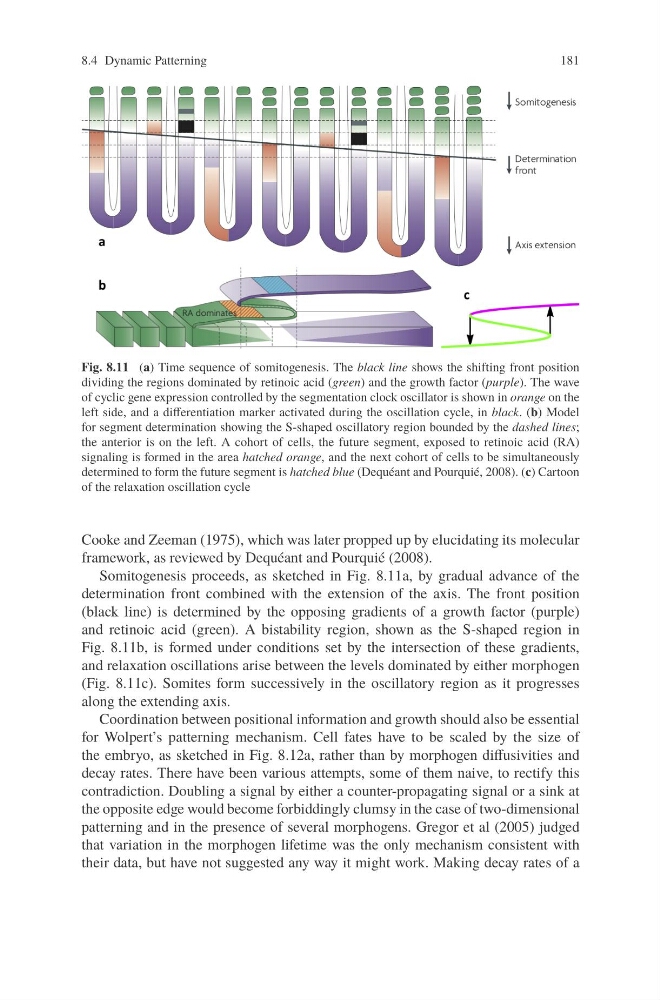
Fig. 8.11 (a) Time sequence of somitogenesis. The black line shows the shifting front position
dividing the regions dominated by retinoic acid (green) and the growth factor (purple). The wave
of cyclic gene expression controlled by the segmentation clock oscillator is shown in orange on the
left side, and a differentiation marker activated during the oscillation cycle, in black. (b) Model
for segment determination showing the S-shaped oscillatory region bounded by the dashed lines;
the anterior is on the left. A cohort of cells, the future segment, exposed to retinoic acid (RA)
signaling is formed in the area hatched orange, and the next cohort of cells to be simultaneously
determined to form the future segment is hatched blue (Dequéant and Pourquié, 2008). (c) Cartoon
of the relaxation oscillation cycle
Cooke and Zeeman (1975), which was later propped up by elucidating its molecular
framework, as reviewed by Dequéant and Pourquié (2008).
Somitogenesis proceeds, as sketched in Fig. 8.11a, by gradual advance of the
determination front combined with the extension of the axis. The front position
(black line) is determined by the opposing gradients of a growth factor (purple)
and retinoic acid (green). A bistability region, shown as the S-shaped region in
Fig. 8.11b, is formed under conditions set by the intersection of these gradients,
and relaxation oscillations arise between the levels dominated by either morphogen
(Fig. 8.11c). Somites form successively in the oscillatory region as it progresses
along the extending axis.
Coordination between positional information and growth should also be essential
for Wolpert’s patterning mechanism. Cell fates have to be scaled by the size of
the embryo, as sketched in Fig. 8.12a, rather than by morphogen diffusivities and
decay rates. There have been various attempts, some of them naive, to rectify this
contradiction. Doubling a signal by either a counter-propagating signal or a sink at
the opposite edge would become forbiddingly clumsy in the case of two-dimensional
patterning and in the presence of several morphogens. Gregor et al (2005) judged
that variation in the morphogen lifetime was the only mechanism consistent with
their data, but have not suggested any way it might work. Making decay rates of a
181
Fig. 8.11 (a) Time sequence of somitogenesis. The black line shows the shifting front position
dividing the regions dominated by retinoic acid (green) and the growth factor (purple). The wave
of cyclic gene expression controlled by the segmentation clock oscillator is shown in orange on the
left side, and a differentiation marker activated during the oscillation cycle, in black. (b) Model
for segment determination showing the S-shaped oscillatory region bounded by the dashed lines;
the anterior is on the left. A cohort of cells, the future segment, exposed to retinoic acid (RA)
signaling is formed in the area hatched orange, and the next cohort of cells to be simultaneously
determined to form the future segment is hatched blue (Dequéant and Pourquié, 2008). (c) Cartoon
of the relaxation oscillation cycle
Cooke and Zeeman (1975), which was later propped up by elucidating its molecular
framework, as reviewed by Dequéant and Pourquié (2008).
Somitogenesis proceeds, as sketched in Fig. 8.11a, by gradual advance of the
determination front combined with the extension of the axis. The front position
(black line) is determined by the opposing gradients of a growth factor (purple)
and retinoic acid (green). A bistability region, shown as the S-shaped region in
Fig. 8.11b, is formed under conditions set by the intersection of these gradients,
and relaxation oscillations arise between the levels dominated by either morphogen
(Fig. 8.11c). Somites form successively in the oscillatory region as it progresses
along the extending axis.
Coordination between positional information and growth should also be essential
for Wolpert’s patterning mechanism. Cell fates have to be scaled by the size of
the embryo, as sketched in Fig. 8.12a, rather than by morphogen diffusivities and
decay rates. There have been various attempts, some of them naive, to rectify this
contradiction. Doubling a signal by either a counter-propagating signal or a sink at
the opposite edge would become forbiddingly clumsy in the case of two-dimensional
patterning and in the presence of several morphogens. Gregor et al (2005) judged
that variation in the morphogen lifetime was the only mechanism consistent with
their data, but have not suggested any way it might work. Making decay rates of a