

8.3 Cell Fates
179
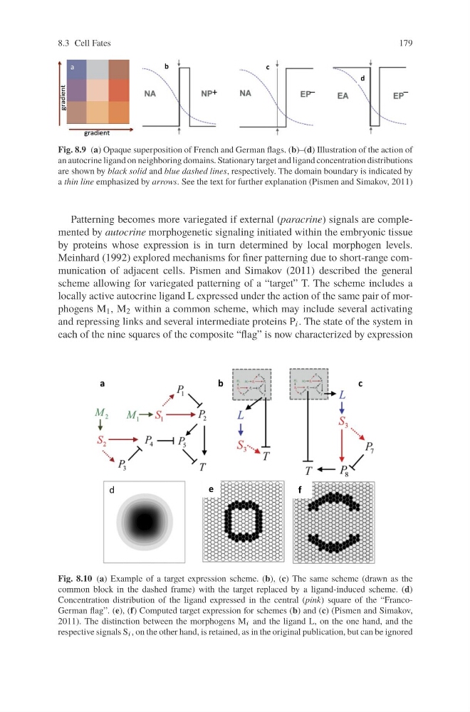
Fig. 8.9 (a) Opaque superposition of French and German flags. (b)–(d) Illustration of the action of
an autocrine ligand on neighboring domains. Stationary target and ligand concentration distributions
are shown by black solid and blue dashed lines, respectively. The domain boundary is indicated by
a thin line emphasized by arrows. See the text for further explanation (Pismen and Simakov, 2011)
Patterning becomes more variegated if external (paracrine) signals are complemented by autocrine morphogenetic signaling initiated within the embryonic tissue
by proteins whose expression is in turn determined by local morphogen levels.
Meinhard (1992) explored mechanisms for finer patterning due to short-range communication of adjacent cells. Pismen and Simakov (2011) described the general
scheme allowing for variegated patterning of a “target” T. The scheme includes a
locally active autocrine ligand L expressed under the action of the same pair of morphogens M 1 , M 2 within a common scheme, which may include several activating
and repressing links and several intermediate proteins P i . The state of the system in
each of the nine squares of the composite “flag” is now characterized by expression
Fig. 8.10 (a) Example of a target expression scheme. (b), (c) The same scheme (drawn as the
common block in the dashed frame) with the target replaced by a ligand-induced scheme. (d)
Concentration distribution of the ligand expressed in the central (pink) square of the “FrancoGerman flag”. (e), (f) Computed target expression for schemes (b) and (c) (Pismen and Simakov,
2011). The distinction between the morphogens M i and the ligand L, on the one hand, and the
respective signals S i , on the other hand, is retained, as in the original publication, but can be ignored
179
Fig. 8.9 (a) Opaque superposition of French and German flags. (b)–(d) Illustration of the action of
an autocrine ligand on neighboring domains. Stationary target and ligand concentration distributions
are shown by black solid and blue dashed lines, respectively. The domain boundary is indicated by
a thin line emphasized by arrows. See the text for further explanation (Pismen and Simakov, 2011)
Patterning becomes more variegated if external (paracrine) signals are complemented by autocrine morphogenetic signaling initiated within the embryonic tissue
by proteins whose expression is in turn determined by local morphogen levels.
Meinhard (1992) explored mechanisms for finer patterning due to short-range communication of adjacent cells. Pismen and Simakov (2011) described the general
scheme allowing for variegated patterning of a “target” T. The scheme includes a
locally active autocrine ligand L expressed under the action of the same pair of morphogens M 1 , M 2 within a common scheme, which may include several activating
and repressing links and several intermediate proteins P i . The state of the system in
each of the nine squares of the composite “flag” is now characterized by expression
Fig. 8.10 (a) Example of a target expression scheme. (b), (c) The same scheme (drawn as the
common block in the dashed frame) with the target replaced by a ligand-induced scheme. (d)
Concentration distribution of the ligand expressed in the central (pink) square of the “FrancoGerman flag”. (e), (f) Computed target expression for schemes (b) and (c) (Pismen and Simakov,
2011). The distinction between the morphogens M i and the ligand L, on the one hand, and the
respective signals S i , on the other hand, is retained, as in the original publication, but can be ignored