

124
6 Active Gels
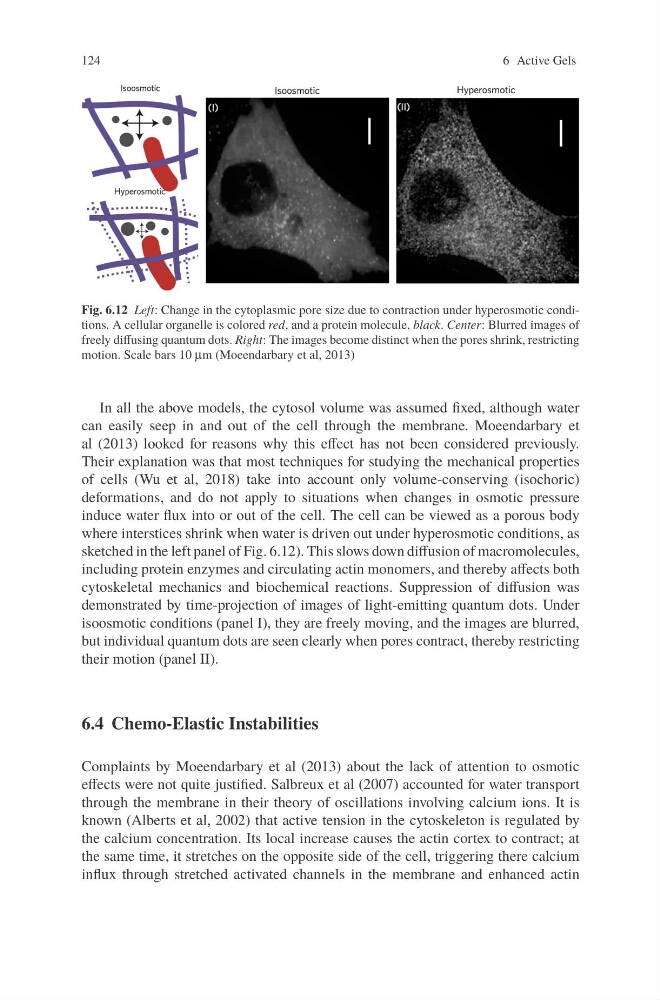
Fig. 6.12 Left: Change in the cytoplasmic pore size due to contraction under hyperosmotic conditions. A cellular organelle is colored red, and a protein molecule, black. Center: Blurred images of
freely diffusing quantum dots. Right: The images become distinct when the pores shrink, restricting
motion. Scale bars 10
In all the above models, the cytosol volume was assumed fixed, although water
can easily seep in and out of the cell through the membrane. Moeendarbary et
al (2013) looked for reasons why this effect has not been considered previously.
Their explanation was that most techniques for studying the mechanical properties
of cells (Wu et al, 2018) take into account only volume-conserving (isochoric)
deformations, and do not apply to situations when changes in osmotic pressure
induce water flux into or out of the cell. The cell can be viewed as a porous body
where interstices shrink when water is driven out under hyperosmotic conditions, as
sketched in the left panel of Fig. 6.12). This slows down diffusion of macromolecules,
including protein enzymes and circulating actin monomers, and thereby affects both
cytoskeletal mechanics and biochemical reactions. Suppression of diffusion was
demonstrated by time-projection of images of light-emitting quantum dots. Under
isoosmotic conditions (panel I), they are freely moving, and the images are blurred,
but individual quantum dots are seen clearly when pores contract, thereby restricting
their motion (panel II).
6.4 Chemo-Elastic Instabilities
Complaints by Moeendarbary et al (2013) about the lack of attention to osmotic
effects were not quite justified. Salbreux et al (2007) accounted for water transport
through the membrane in their theory of oscillations involving calcium ions. It is
known (Alberts et al, 2002) that active tension in the cytoskeleton is regulated by
the calcium concentration. Its local increase causes the actin cortex to contract; at
the same time, it stretches on the opposite side of the cell, triggering there calcium
influx through stretched activated channels in the membrane and enhanced actin
(Moeendarbary et al, 2013)
μm
6 Active Gels
Fig. 6.12 Left: Change in the cytoplasmic pore size due to contraction under hyperosmotic conditions. A cellular organelle is colored red, and a protein molecule, black. Center: Blurred images of
freely diffusing quantum dots. Right: The images become distinct when the pores shrink, restricting
motion. Scale bars 10
In all the above models, the cytosol volume was assumed fixed, although water
can easily seep in and out of the cell through the membrane. Moeendarbary et
al (2013) looked for reasons why this effect has not been considered previously.
Their explanation was that most techniques for studying the mechanical properties
of cells (Wu et al, 2018) take into account only volume-conserving (isochoric)
deformations, and do not apply to situations when changes in osmotic pressure
induce water flux into or out of the cell. The cell can be viewed as a porous body
where interstices shrink when water is driven out under hyperosmotic conditions, as
sketched in the left panel of Fig. 6.12). This slows down diffusion of macromolecules,
including protein enzymes and circulating actin monomers, and thereby affects both
cytoskeletal mechanics and biochemical reactions. Suppression of diffusion was
demonstrated by time-projection of images of light-emitting quantum dots. Under
isoosmotic conditions (panel I), they are freely moving, and the images are blurred,
but individual quantum dots are seen clearly when pores contract, thereby restricting
their motion (panel II).
6.4 Chemo-Elastic Instabilities
Complaints by Moeendarbary et al (2013) about the lack of attention to osmotic
effects were not quite justified. Salbreux et al (2007) accounted for water transport
through the membrane in their theory of oscillations involving calcium ions. It is
known (Alberts et al, 2002) that active tension in the cytoskeleton is regulated by
the calcium concentration. Its local increase causes the actin cortex to contract; at
the same time, it stretches on the opposite side of the cell, triggering there calcium
influx through stretched activated channels in the membrane and enhanced actin
(Moeendarbary et al, 2013)
μm