

92
5 Eukaryotic Cells
sapiens genome codes proteins taking part in the assembly of actin filaments alone.
Researchers care no less about it, uncovering minute details from the molecular to
the mechanical level.
Armies of biologists, biochemists, and biophysicists, more generously funded than
their colleagues in any other branch of science, study various modes of operation of
the cells in the innards of healthy or sick individuals or along the conveyor belt of
organism development. Thanks to these studies, we live on the average some twenty
years longer than hundred years ago, but we still know much less than we don’t know.
In this book, we cannot delve deeply into the workings of the cell. Its intricacies are
hidden in its chemistry, but for us, here as in diffusiophoresis or bacterial growth,
chemistry provides an unspecified source of energy driving the mechanics of an
active medium. Yet, we need to view this medium here in finer detail than up to
now. Even though we restrict in this chapter to mechanical aspects of the cell, we
arrive at the peak of complexity midway through the book, based on observations
and experiment rather than theories and simulations.
5.2 Filaments and Motors
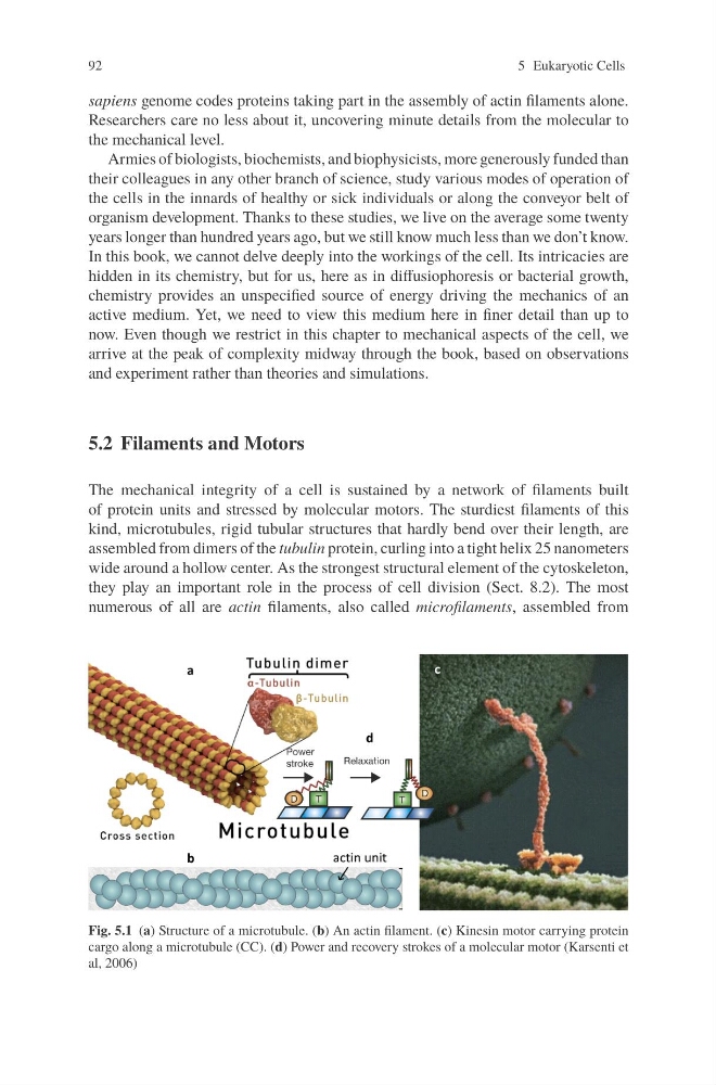
The mechanical integrity of a cell is sustained by a network of filaments built
of protein units and stressed by molecular motors. The sturdiest filaments of this
kind, microtubules, rigid tubular structures that hardly bend over their length, are
assembled from dimers of the tubulin protein, curling into a tight helix 25 nanometers
wide around a hollow center. As the strongest structural element of the cytoskeleton,
they play an important role in the process of cell division (Sect. 8.2). The most
numerous of all are actin filaments, also called microfilaments, assembled from
Fig. 5.1 (a) Structure of a microtubule. (b) An actin filament. (c) Kinesin motor carrying protein
cargo along a microtubule (CC). (d) Power and recovery strokes of a molecular motor (Karsenti et
al, 2006)
5 Eukaryotic Cells
sapiens genome codes proteins taking part in the assembly of actin filaments alone.
Researchers care no less about it, uncovering minute details from the molecular to
the mechanical level.
Armies of biologists, biochemists, and biophysicists, more generously funded than
their colleagues in any other branch of science, study various modes of operation of
the cells in the innards of healthy or sick individuals or along the conveyor belt of
organism development. Thanks to these studies, we live on the average some twenty
years longer than hundred years ago, but we still know much less than we don’t know.
In this book, we cannot delve deeply into the workings of the cell. Its intricacies are
hidden in its chemistry, but for us, here as in diffusiophoresis or bacterial growth,
chemistry provides an unspecified source of energy driving the mechanics of an
active medium. Yet, we need to view this medium here in finer detail than up to
now. Even though we restrict in this chapter to mechanical aspects of the cell, we
arrive at the peak of complexity midway through the book, based on observations
and experiment rather than theories and simulations.
5.2 Filaments and Motors
The mechanical integrity of a cell is sustained by a network of filaments built
of protein units and stressed by molecular motors. The sturdiest filaments of this
kind, microtubules, rigid tubular structures that hardly bend over their length, are
assembled from dimers of the tubulin protein, curling into a tight helix 25 nanometers
wide around a hollow center. As the strongest structural element of the cytoskeleton,
they play an important role in the process of cell division (Sect. 8.2). The most
numerous of all are actin filaments, also called microfilaments, assembled from
Fig. 5.1 (a) Structure of a microtubule. (b) An actin filament. (c) Kinesin motor carrying protein
cargo along a microtubule (CC). (d) Power and recovery strokes of a molecular motor (Karsenti et
al, 2006)