

278
W. Qiu and X.-Y. Liu
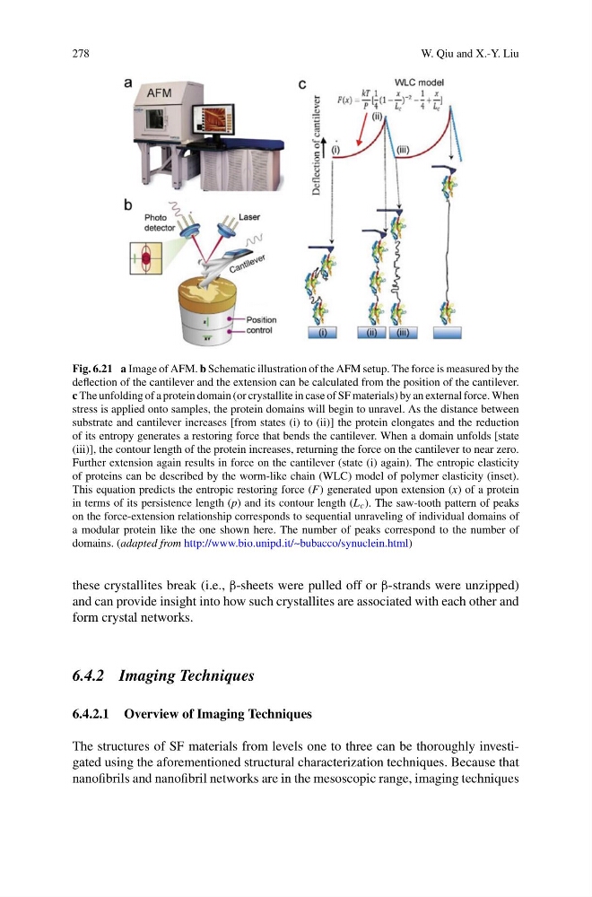
Fig. 6.21 a Image of AFM. b Schematic illustration of the AFM setup. The force is measured by the
deflection of the cantilever and the extension can be calculated from the position of the cantilever.
c The unfolding of a protein domain (or crystallite in case of SF materials) by an external force. When
stress is applied onto samples, the protein domains will begin to unravel. As the distance between
substrate and cantilever increases [from states (i) to (ii)] the protein elongates and the reduction
of its entropy generates a restoring force that bends the cantilever. When a domain unfolds [state
(iii)], the contour length of the protein increases, returning the force on the cantilever to near zero.
Further extension again results in force on the cantilever (state (i) again). The entropic elasticity
of proteins can be described by the worm-like chain (WLC) model of polymer elasticity (inset).
This equation predicts the entropic restoring force (F) generated upon extension (x) of a protein
in terms of its persistence length (p) and its contour length (L c ). The saw-tooth pattern of peaks
on the force-extension relationship corresponds to sequential unraveling of individual domains of
a modular protein like the one shown here. The number of peaks correspond to the number of
domains. (adapted from http://www.bio.unipd.it/~bubacco/synuclein.html)
these crystallites break (i.e., β-sheets were pulled off or β-strands were unzipped)
and can provide insight into how such crystallites are associated with each other and
form crystal networks.
6.4.2 Imaging Techniques
6.4.2.1 Overview of Imaging Techniques
The structures of SF materials from levels one to three can be thoroughly investigated using the aforementioned structural characterization techniques. Because that
nanofibrils and nanofibril networks are in the mesoscopic range, imaging techniques
W. Qiu and X.-Y. Liu
Fig. 6.21 a Image of AFM. b Schematic illustration of the AFM setup. The force is measured by the
deflection of the cantilever and the extension can be calculated from the position of the cantilever.
c The unfolding of a protein domain (or crystallite in case of SF materials) by an external force. When
stress is applied onto samples, the protein domains will begin to unravel. As the distance between
substrate and cantilever increases [from states (i) to (ii)] the protein elongates and the reduction
of its entropy generates a restoring force that bends the cantilever. When a domain unfolds [state
(iii)], the contour length of the protein increases, returning the force on the cantilever to near zero.
Further extension again results in force on the cantilever (state (i) again). The entropic elasticity
of proteins can be described by the worm-like chain (WLC) model of polymer elasticity (inset).
This equation predicts the entropic restoring force (F) generated upon extension (x) of a protein
in terms of its persistence length (p) and its contour length (L c ). The saw-tooth pattern of peaks
on the force-extension relationship corresponds to sequential unraveling of individual domains of
a modular protein like the one shown here. The number of peaks correspond to the number of
domains. (adapted from http://www.bio.unipd.it/~bubacco/synuclein.html)
these crystallites break (i.e., β-sheets were pulled off or β-strands were unzipped)
and can provide insight into how such crystallites are associated with each other and
form crystal networks.
6.4.2 Imaging Techniques
6.4.2.1 Overview of Imaging Techniques
The structures of SF materials from levels one to three can be thoroughly investigated using the aforementioned structural characterization techniques. Because that
nanofibrils and nanofibril networks are in the mesoscopic range, imaging techniques