

274
W. Qiu and X.-Y. Liu
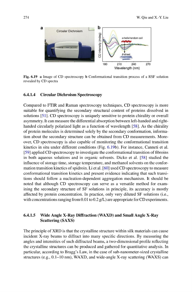
Fig. 6.19 a Image of CD spectroscopy b Conformational transition process of a RSF solution
revealed by CD spectra
6.4.1.4 Circular Dichroism Spectroscopy
Compared to FTIR and Raman spectroscopy techniques, CD spectroscopy is more
suitable for quantifying the secondary structural content of proteins dissolved in
solutions [51]. CD spectroscopy is uniquely sensitive to protein chirality or overall
asymmetry. It can measure the differential absorption between left-handed and righthanded circularly polarized light as a function of wavelength [58]. As the chirality
of protein molecules is determined solely by the secondary conformation, information about the secondary structure can be obtained from CD measurements. Moreover, CD spectroscopy is also capable of monitoring the conformational transition
kinetics in situ under different conditions (Fig. 6.19b). For instance, Canneti et al.
[59] applied CD spectroscopy to investigate the conformational transition of fibroins
in both aqueous solutions and in organic solvents. Dicko et al. [58] studied the
influence of storage time, storage temperature, and methanol solvents on the conformation transition kinetics of spidroin. Li et al. [60] used CD spectroscopy to measure
conformational transition kinetics and present evidence indicating that such transitions should follow a nucleation-dependent aggregation mechanism. It should be
noted that although CD spectroscopy can serve as a versatile method for examining the secondary structure of SF solutions in principle, its accuracy is mostly
affected by protein concentration. In practice, only very diluted SF solutions (i.e.,
with concentrations ranging from 0.01 to 0.2 g/L) are appropriate for CD experiments.
6.4.1.5 Wide Angle X-Ray Diffraction (WAXD) and Small Angle X-Ray
Scattering (SAXS)
The principle of XRD is that the crystalline structure within silk materials can cause
incident X-ray beams to diffract into many specific directions. By measuring the
angles and intensities of such diffracted beams, a two-dimensional profile reflecting
the crystalline structures can be produced and gathered for quantitative analysis. In
particular, according to Bragg’s Law, in the case of sub-nanometer-sized crystalline
structures (e.g., 0.1–10 nm), WAXD, and wide-angle X-ray scattering (WAXS) can
W. Qiu and X.-Y. Liu
Fig. 6.19 a Image of CD spectroscopy b Conformational transition process of a RSF solution
revealed by CD spectra
6.4.1.4 Circular Dichroism Spectroscopy
Compared to FTIR and Raman spectroscopy techniques, CD spectroscopy is more
suitable for quantifying the secondary structural content of proteins dissolved in
solutions [51]. CD spectroscopy is uniquely sensitive to protein chirality or overall
asymmetry. It can measure the differential absorption between left-handed and righthanded circularly polarized light as a function of wavelength [58]. As the chirality
of protein molecules is determined solely by the secondary conformation, information about the secondary structure can be obtained from CD measurements. Moreover, CD spectroscopy is also capable of monitoring the conformational transition
kinetics in situ under different conditions (Fig. 6.19b). For instance, Canneti et al.
[59] applied CD spectroscopy to investigate the conformational transition of fibroins
in both aqueous solutions and in organic solvents. Dicko et al. [58] studied the
influence of storage time, storage temperature, and methanol solvents on the conformation transition kinetics of spidroin. Li et al. [60] used CD spectroscopy to measure
conformational transition kinetics and present evidence indicating that such transitions should follow a nucleation-dependent aggregation mechanism. It should be
noted that although CD spectroscopy can serve as a versatile method for examining the secondary structure of SF solutions in principle, its accuracy is mostly
affected by protein concentration. In practice, only very diluted SF solutions (i.e.,
with concentrations ranging from 0.01 to 0.2 g/L) are appropriate for CD experiments.
6.4.1.5 Wide Angle X-Ray Diffraction (WAXD) and Small Angle X-Ray
Scattering (SAXS)
The principle of XRD is that the crystalline structure within silk materials can cause
incident X-ray beams to diffract into many specific directions. By measuring the
angles and intensities of such diffracted beams, a two-dimensional profile reflecting
the crystalline structures can be produced and gathered for quantitative analysis. In
particular, according to Bragg’s Law, in the case of sub-nanometer-sized crystalline
structures (e.g., 0.1–10 nm), WAXD, and wide-angle X-ray scattering (WAXS) can