

376
P. Martínez-Vázquez et al.
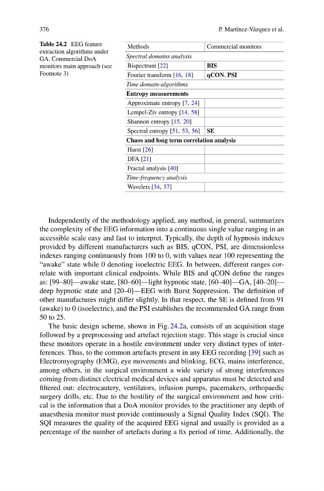
Table 24.2 EEG feature
extraction algorithms under
GA. Commercial DoA
monitors main approach (see
Footnote 3)
Methods
Commercial monitors
Spectral domains analysis
Bispectrum [22]
BIS
Fourier transform [16, 18]
qCON, PSI
Time domain-algorithms
Entropy measurements
Approximate entropy [7, 24]
Lempel-Ziv entropy [14, 58]
Shannon entropy [15, 20]
Spectral entropy [51, 53, 56] SE
Chaos and long term correlation analysis
Hurst [26]
DFA [21]
Fractal analysis [40]
Time-frequency analysis
Wavelets [34, 37]
Independently of the methodology applied, any method, in general, summarizes
the complexity of the EEG information into a continuous single value ranging in an
accessible scale easy and fast to interpret. Typically, the depth of hypnosis indexes
provided by different manufacturers such as BIS, qCON, PSI, are dimensionless
indexes ranging continuously from 100 to 0, with values near 100 representing the
“awake” state while 0 denoting isoelectric EEG. In between, different ranges correlate with important clinical endpoints. While BIS and qCON define the ranges
as: [99–80]—awake state, [80–60]—light hypnotic state, [60–40]—GA, [40–20]—
deep hypnotic state and [20–0]—EEG with Burst Suppression. The definition of
other manufactures might differ slightly. In that respect, the SE is defined from 91
(awake) to 0 (isoelectric), and the PSI establishes the recommended GA range from
50 to 25.
The basic design scheme, shown in Fig. 24.2a, consists of an acquisition stage
followed by a preprocessing and artefact rejection stage. This stage is crucial since
these monitors operate in a hostile environment under very distinct types of interferences. Thus, to the common artefacts present in any EEG recording [39] such as
Electromyography (EMG), eye movements and blinking, ECG, mains interference,
among others, in the surgical environment a wide variety of strong interferences
coming from distinct electrical medical devices and apparatus must be detected and
filtered out: electrocautery, ventilators, infusion pumps, pacemakers, orthopaedic
surgery drills, etc. Due to the hostility of the surgical environment and how critical is the information that a DoA monitor provides to the practitioner any depth of
anaesthesia monitor must provide continuously a Signal Quality Index (SQI). The
SQI measures the quality of the acquired EEG signal and usually is provided as a
percentage of the number of artefacts during a fix period of time. Additionally, the
P. Martínez-Vázquez et al.
Table 24.2 EEG feature
extraction algorithms under
GA. Commercial DoA
monitors main approach (see
Footnote 3)
Methods
Commercial monitors
Spectral domains analysis
Bispectrum [22]
BIS
Fourier transform [16, 18]
qCON, PSI
Time domain-algorithms
Entropy measurements
Approximate entropy [7, 24]
Lempel-Ziv entropy [14, 58]
Shannon entropy [15, 20]
Spectral entropy [51, 53, 56] SE
Chaos and long term correlation analysis
Hurst [26]
DFA [21]
Fractal analysis [40]
Time-frequency analysis
Wavelets [34, 37]
Independently of the methodology applied, any method, in general, summarizes
the complexity of the EEG information into a continuous single value ranging in an
accessible scale easy and fast to interpret. Typically, the depth of hypnosis indexes
provided by different manufacturers such as BIS, qCON, PSI, are dimensionless
indexes ranging continuously from 100 to 0, with values near 100 representing the
“awake” state while 0 denoting isoelectric EEG. In between, different ranges correlate with important clinical endpoints. While BIS and qCON define the ranges
as: [99–80]—awake state, [80–60]—light hypnotic state, [60–40]—GA, [40–20]—
deep hypnotic state and [20–0]—EEG with Burst Suppression. The definition of
other manufactures might differ slightly. In that respect, the SE is defined from 91
(awake) to 0 (isoelectric), and the PSI establishes the recommended GA range from
50 to 25.
The basic design scheme, shown in Fig. 24.2a, consists of an acquisition stage
followed by a preprocessing and artefact rejection stage. This stage is crucial since
these monitors operate in a hostile environment under very distinct types of interferences. Thus, to the common artefacts present in any EEG recording [39] such as
Electromyography (EMG), eye movements and blinking, ECG, mains interference,
among others, in the surgical environment a wide variety of strong interferences
coming from distinct electrical medical devices and apparatus must be detected and
filtered out: electrocautery, ventilators, infusion pumps, pacemakers, orthopaedic
surgery drills, etc. Due to the hostility of the surgical environment and how critical is the information that a DoA monitor provides to the practitioner any depth of
anaesthesia monitor must provide continuously a Signal Quality Index (SQI). The
SQI measures the quality of the acquired EEG signal and usually is provided as a
percentage of the number of artefacts during a fix period of time. Additionally, the