

23 General Anaesthesia and Oscillations in Human Physiology …
367
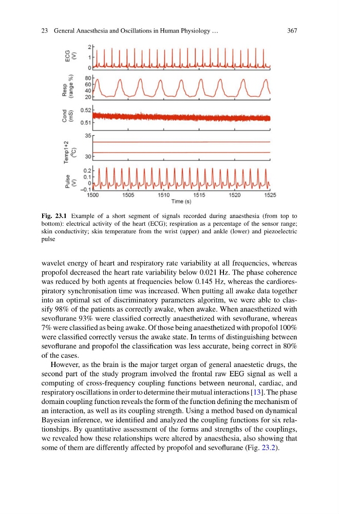
Fig. 23.1 Example of a short segment of signals recorded during anaesthesia (from top to
bottom): electrical activity of the heart (ECG); respiration as a percentage of the sensor range;
skin conductivity; skin temperature from the wrist (upper) and ankle (lower) and piezoelectric
pulse
wavelet energy of heart and respiratory rate variability at all frequencies, whereas
propofol decreased the heart rate variability below 0.021 Hz. The phase coherence
was reduced by both agents at frequencies below 0.145 Hz, whereas the cardiorespiratory synchronisation time was increased. When putting all awake data together
into an optimal set of discriminatory parameters algoritm, we were able to classify 98% of the patients as correctly awake, when awake. When anaesthetized with
sevoflurane 93% were classified correctly anaesthetized with sevoflurane, whereas
7% were classified as being awake. Of those being anaesthetized with propofol 100%
were classified correctly versus the awake state. In terms of distinguishing between
sevoflurane and propofol the classification was less accurate, being correct in 80%
of the cases.
However, as the brain is the major target organ of general anaestetic drugs, the
second part of the study program involved the frontal raw EEG signal as well a
computing of cross-frequency coupling functions between neuronal, cardiac, and
respiratory oscillations in order to determine their mutual interactions [13]. The phase
domain coupling function reveals the form of the function defining the mechanism of
an interaction, as well as its coupling strength. Using a method based on dynamical
Bayesian inference, we identified and analyzed the coupling functions for six relationships. By quantitative assessment of the forms and strengths of the couplings,
we revealed how these relationships were altered by anaesthesia, also showing that
some of them are differently affected by propofol and sevoflurane (Fig. 23.2).
367
Fig. 23.1 Example of a short segment of signals recorded during anaesthesia (from top to
bottom): electrical activity of the heart (ECG); respiration as a percentage of the sensor range;
skin conductivity; skin temperature from the wrist (upper) and ankle (lower) and piezoelectric
pulse
wavelet energy of heart and respiratory rate variability at all frequencies, whereas
propofol decreased the heart rate variability below 0.021 Hz. The phase coherence
was reduced by both agents at frequencies below 0.145 Hz, whereas the cardiorespiratory synchronisation time was increased. When putting all awake data together
into an optimal set of discriminatory parameters algoritm, we were able to classify 98% of the patients as correctly awake, when awake. When anaesthetized with
sevoflurane 93% were classified correctly anaesthetized with sevoflurane, whereas
7% were classified as being awake. Of those being anaesthetized with propofol 100%
were classified correctly versus the awake state. In terms of distinguishing between
sevoflurane and propofol the classification was less accurate, being correct in 80%
of the cases.
However, as the brain is the major target organ of general anaestetic drugs, the
second part of the study program involved the frontal raw EEG signal as well a
computing of cross-frequency coupling functions between neuronal, cardiac, and
respiratory oscillations in order to determine their mutual interactions [13]. The phase
domain coupling function reveals the form of the function defining the mechanism of
an interaction, as well as its coupling strength. Using a method based on dynamical
Bayesian inference, we identified and analyzed the coupling functions for six relationships. By quantitative assessment of the forms and strengths of the couplings,
we revealed how these relationships were altered by anaesthesia, also showing that
some of them are differently affected by propofol and sevoflurane (Fig. 23.2).