

20 Sleep-Related Modulations of Heart Rate Variability…
315
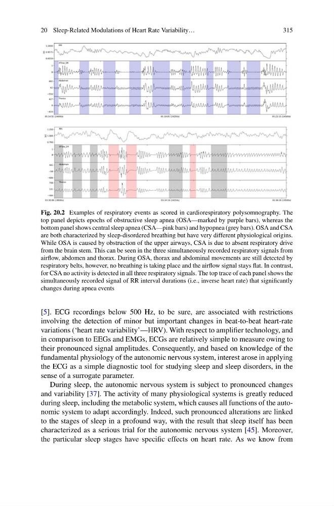
Fig. 20.2 Examples of respiratory events as scored in cardiorespiratory polysomnography. The
top panel depicts epochs of obstructive sleep apnea (OSA—marked by purple bars), whereas the
bottom panel shows central sleep apnea (CSA—pink bars) and hypopnea (grey bars). OSA and CSA
are both characterized by sleep-disordered breathing but have very different physiological origins.
While OSA is caused by obstruction of the upper airways, CSA is due to absent respiratory drive
from the brain stem. This can be seen in the three simultaneously recorded respiratory signals from
airflow, abdomen and thorax. During OSA, thorax and abdominal movements are still detected by
respiratory belts, however, no breathing is taking place and the airflow signal stays flat. In contrast,
for CSA no activity is detected in all three respiratory signals. The top trace of each panel shows the
simultaneously recorded signal of RR interval durations (i.e., inverse heart rate) that significantly
changes during apnea events
[5]. ECG recordings below 500 Hz, to be sure, are associated with restrictions
involving the detection of minor but important changes in beat-to-beat heart-rate
variations (‘heart rate variability’—HRV). With respect to amplifier technology, and
in comparison to EEGs and EMGs, ECGs are relatively simple to measure owing to
their pronounced signal amplitudes. Consequently, and based on knowledge of the
fundamental physiology of the autonomic nervous system, interest arose in applying
the ECG as a simple diagnostic tool for studying sleep and sleep disorders, in the
sense of a surrogate parameter.
During sleep, the autonomic nervous system is subject to pronounced changes
and variability [37]. The activity of many physiological systems is greatly reduced
during sleep, including the metabolic system, which causes all functions of the autonomic system to adapt accordingly. Indeed, such pronounced alterations are linked
to the stages of sleep in a profound way, with the result that sleep itself has been
characterized as a serious trial for the autonomic nervous system [45]. Moreover,
the particular sleep stages have specific effects on heart rate. As we know from
315
Fig. 20.2 Examples of respiratory events as scored in cardiorespiratory polysomnography. The
top panel depicts epochs of obstructive sleep apnea (OSA—marked by purple bars), whereas the
bottom panel shows central sleep apnea (CSA—pink bars) and hypopnea (grey bars). OSA and CSA
are both characterized by sleep-disordered breathing but have very different physiological origins.
While OSA is caused by obstruction of the upper airways, CSA is due to absent respiratory drive
from the brain stem. This can be seen in the three simultaneously recorded respiratory signals from
airflow, abdomen and thorax. During OSA, thorax and abdominal movements are still detected by
respiratory belts, however, no breathing is taking place and the airflow signal stays flat. In contrast,
for CSA no activity is detected in all three respiratory signals. The top trace of each panel shows the
simultaneously recorded signal of RR interval durations (i.e., inverse heart rate) that significantly
changes during apnea events
[5]. ECG recordings below 500 Hz, to be sure, are associated with restrictions
involving the detection of minor but important changes in beat-to-beat heart-rate
variations (‘heart rate variability’—HRV). With respect to amplifier technology, and
in comparison to EEGs and EMGs, ECGs are relatively simple to measure owing to
their pronounced signal amplitudes. Consequently, and based on knowledge of the
fundamental physiology of the autonomic nervous system, interest arose in applying
the ECG as a simple diagnostic tool for studying sleep and sleep disorders, in the
sense of a surrogate parameter.
During sleep, the autonomic nervous system is subject to pronounced changes
and variability [37]. The activity of many physiological systems is greatly reduced
during sleep, including the metabolic system, which causes all functions of the autonomic system to adapt accordingly. Indeed, such pronounced alterations are linked
to the stages of sleep in a profound way, with the result that sleep itself has been
characterized as a serious trial for the autonomic nervous system [45]. Moreover,
the particular sleep stages have specific effects on heart rate. As we know from