

278
A. Colantuoni and D. Lapi
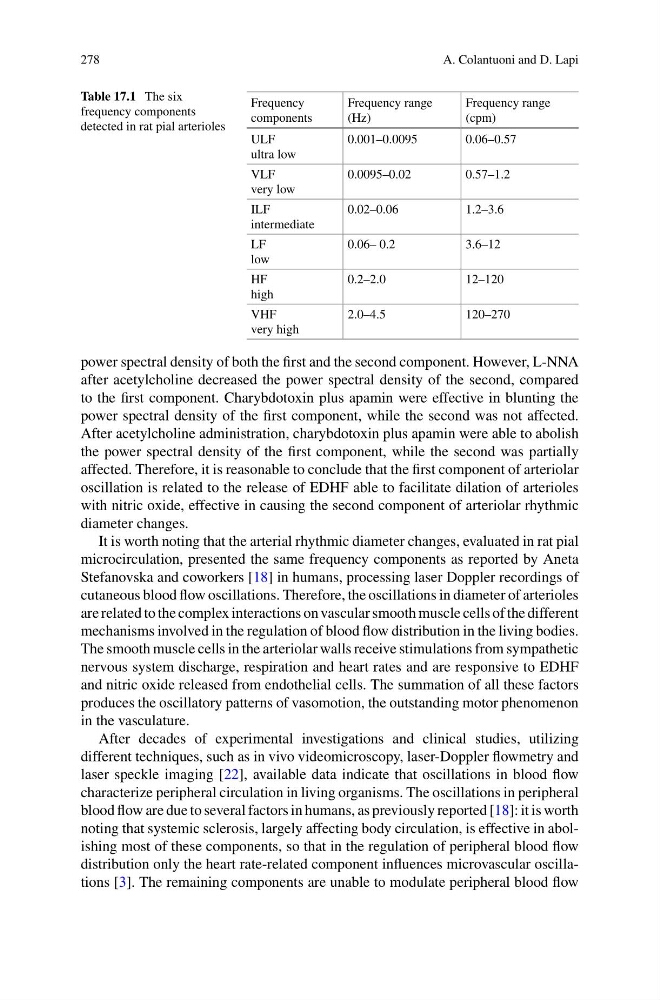
Table 17.1 The six
frequency components
detected in rat pial arterioles
Frequency
components
Frequency range
(Hz)
Frequency range
(cpm)
ULF
ultra low
0.001–0.0095
0.06–0.57
VLF
very low
0.0095–0.02
0.57–1.2
ILF
intermediate
0.02–0.06
1.2–3.6
LF
low
0.06– 0.2
3.6–12
HF
high
0.2–2.0
12–120
VHF
very high
2.0–4.5
120–270
power spectral density of both the first and the second component. However, L-NNA
after acetylcholine decreased the power spectral density of the second, compared
to the first component. Charybdotoxin plus apamin were effective in blunting the
power spectral density of the first component, while the second was not affected.
After acetylcholine administration, charybdotoxin plus apamin were able to abolish
the power spectral density of the first component, while the second was partially
affected. Therefore, it is reasonable to conclude that the first component of arteriolar
oscillation is related to the release of EDHF able to facilitate dilation of arterioles
with nitric oxide, effective in causing the second component of arteriolar rhythmic
diameter changes.
It is worth noting that the arterial rhythmic diameter changes, evaluated in rat pial
microcirculation, presented the same frequency components as reported by Aneta
Stefanovska and coworkers [18] in humans, processing laser Doppler recordings of
cutaneous blood flow oscillations. Therefore, the oscillations in diameter of arterioles
are related to the complex interactions on vascular smooth muscle cells of the different
mechanisms involved in the regulation of blood flow distribution in the living bodies.
The smooth muscle cells in the arteriolar walls receive stimulations from sympathetic
nervous system discharge, respiration and heart rates and are responsive to EDHF
and nitric oxide released from endothelial cells. The summation of all these factors
produces the oscillatory patterns of vasomotion, the outstanding motor phenomenon
in the vasculature.
After decades of experimental investigations and clinical studies, utilizing
different techniques, such as in vivo videomicroscopy, laser-Doppler flowmetry and
laser speckle imaging [22], available data indicate that oscillations in blood flow
characterize peripheral circulation in living organisms. The oscillations in peripheral
blood flow are due to several factors in humans, as previously reported [18]: it is worth
noting that systemic sclerosis, largely affecting body circulation, is effective in abolishing most of these components, so that in the regulation of peripheral blood flow
distribution only the heart rate-related component influences microvascular oscillations [3]. The remaining components are unable to modulate peripheral blood flow
A. Colantuoni and D. Lapi
Table 17.1 The six
frequency components
detected in rat pial arterioles
Frequency
components
Frequency range
(Hz)
Frequency range
(cpm)
ULF
ultra low
0.001–0.0095
0.06–0.57
VLF
very low
0.0095–0.02
0.57–1.2
ILF
intermediate
0.02–0.06
1.2–3.6
LF
low
0.06– 0.2
3.6–12
HF
high
0.2–2.0
12–120
VHF
very high
2.0–4.5
120–270
power spectral density of both the first and the second component. However, L-NNA
after acetylcholine decreased the power spectral density of the second, compared
to the first component. Charybdotoxin plus apamin were effective in blunting the
power spectral density of the first component, while the second was not affected.
After acetylcholine administration, charybdotoxin plus apamin were able to abolish
the power spectral density of the first component, while the second was partially
affected. Therefore, it is reasonable to conclude that the first component of arteriolar
oscillation is related to the release of EDHF able to facilitate dilation of arterioles
with nitric oxide, effective in causing the second component of arteriolar rhythmic
diameter changes.
It is worth noting that the arterial rhythmic diameter changes, evaluated in rat pial
microcirculation, presented the same frequency components as reported by Aneta
Stefanovska and coworkers [18] in humans, processing laser Doppler recordings of
cutaneous blood flow oscillations. Therefore, the oscillations in diameter of arterioles
are related to the complex interactions on vascular smooth muscle cells of the different
mechanisms involved in the regulation of blood flow distribution in the living bodies.
The smooth muscle cells in the arteriolar walls receive stimulations from sympathetic
nervous system discharge, respiration and heart rates and are responsive to EDHF
and nitric oxide released from endothelial cells. The summation of all these factors
produces the oscillatory patterns of vasomotion, the outstanding motor phenomenon
in the vasculature.
After decades of experimental investigations and clinical studies, utilizing
different techniques, such as in vivo videomicroscopy, laser-Doppler flowmetry and
laser speckle imaging [22], available data indicate that oscillations in blood flow
characterize peripheral circulation in living organisms. The oscillations in peripheral
blood flow are due to several factors in humans, as previously reported [18]: it is worth
noting that systemic sclerosis, largely affecting body circulation, is effective in abolishing most of these components, so that in the regulation of peripheral blood flow
distribution only the heart rate-related component influences microvascular oscillations [3]. The remaining components are unable to modulate peripheral blood flow