

516
V. S. Simi et al.
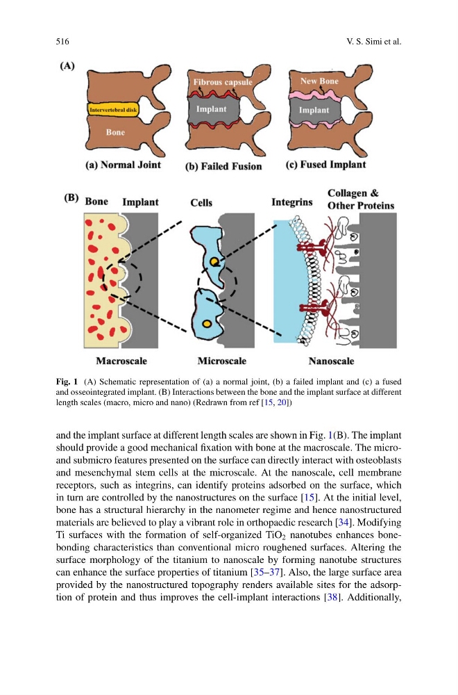
Fig. 1 (A) Schematic representation of (a) a normal joint, (b) a failed implant and (c) a fused
and osseointegrated implant. (B) Interactions between the bone and the implant surface at different
length scales (macro, micro and nano) (Redrawn from ref [15, 20])
and the implant surface at different length scales are shown in Fig. 1(B). The implant
should provide a good mechanical fixation with bone at the macroscale. The microand submicro features presented on the surface can directly interact with osteoblasts
and mesenchymal stem cells at the microscale. At the nanoscale, cell membrane
receptors, such as integrins, can identify proteins adsorbed on the surface, which
in turn are controlled by the nanostructures on the surface [15]. At the initial level,
bone has a structural hierarchy in the nanometer regime and hence nanostructured
materials are believed to play a vibrant role in orthopaedic research [34]. Modifying
Ti surfaces with the formation of self-organized TiO 2 nanotubes enhances bonebonding characteristics than conventional micro roughened surfaces. Altering the
surface morphology of the titanium to nanoscale by forming nanotube structures
can enhance the surface properties of titanium [35–37]. Also, the large surface area
provided by the nanostructured topography renders available sites for the adsorption of protein and thus improves the cell-implant interactions [38]. Additionally,
V. S. Simi et al.
Fig. 1 (A) Schematic representation of (a) a normal joint, (b) a failed implant and (c) a fused
and osseointegrated implant. (B) Interactions between the bone and the implant surface at different
length scales (macro, micro and nano) (Redrawn from ref [15, 20])
and the implant surface at different length scales are shown in Fig. 1(B). The implant
should provide a good mechanical fixation with bone at the macroscale. The microand submicro features presented on the surface can directly interact with osteoblasts
and mesenchymal stem cells at the microscale. At the nanoscale, cell membrane
receptors, such as integrins, can identify proteins adsorbed on the surface, which
in turn are controlled by the nanostructures on the surface [15]. At the initial level,
bone has a structural hierarchy in the nanometer regime and hence nanostructured
materials are believed to play a vibrant role in orthopaedic research [34]. Modifying
Ti surfaces with the formation of self-organized TiO 2 nanotubes enhances bonebonding characteristics than conventional micro roughened surfaces. Altering the
surface morphology of the titanium to nanoscale by forming nanotube structures
can enhance the surface properties of titanium [35–37]. Also, the large surface area
provided by the nanostructured topography renders available sites for the adsorption of protein and thus improves the cell-implant interactions [38]. Additionally,