

372
N. Ashwin Kumar et al.
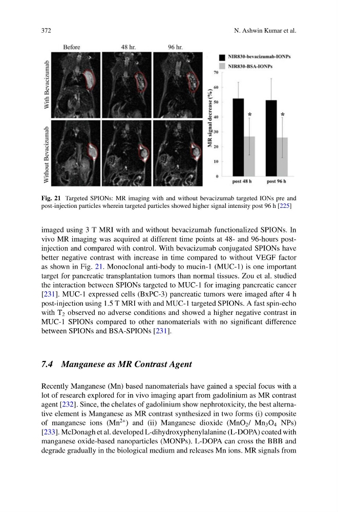
Fig. 21 Targeted SPIONs: MR imaging with and without bevacizumab targeted IONs pre and
post-injection particles wherein targeted particles showed higher signal intensity post 96 h [225]
imaged using 3 T MRI with and without bevacizumab functionalized SPIONs. In
vivo MR imaging was acquired at different time points at 48- and 96-hours postinjection and compared with control. With bevacizumab conjugated SPIONs have
better negative contrast with increase in time compared to without VEGF factor
as shown in Fig. 21. Monoclonal anti-body to mucin-1 (MUC-1) is one important
target for pancreatic transplantation tumors than normal tissues. Zou et al. studied
the interaction between SPIONs targeted to MUC-1 for imaging pancreatic cancer
[231]. MUC-1 expressed cells (BxPC-3) pancreatic tumors were imaged after 4 h
post-injection using 1.5 T MRI with and MUC-1 targeted SPIONs. A fast spin-echo
with T 2 observed no adverse conditions and showed a higher negative contrast in
MUC-1 SPIONs compared to other nanomaterials with no significant difference
between SPIONs and BSA-SPIONs [231].
7.4 Manganese as MR Contrast Agent
Recently Manganese (Mn) based nanomaterials have gained a special focus with a
lot of research explored for in vivo imaging apart from gadolinium as MR contrast
agent [232]. Since, the chelates of gadolinium show nephrotoxicity, the best alternative element is Manganese as MR contrast synthesized in two forms (i) composite
of manganese ions (Mn
2+ ) and (ii) Manganese dioxide (MnO 2 / Mn 3 O 4 NPs)
[233]. McDonagh et al. developed L-dihydroxyphenylalanine (L-DOPA) coated with
manganese oxide-based nanoparticles (MONPs). L-DOPA can cross the BBB and
degrade gradually in the biological medium and releases Mn ions. MR signals from
N. Ashwin Kumar et al.
Fig. 21 Targeted SPIONs: MR imaging with and without bevacizumab targeted IONs pre and
post-injection particles wherein targeted particles showed higher signal intensity post 96 h [225]
imaged using 3 T MRI with and without bevacizumab functionalized SPIONs. In
vivo MR imaging was acquired at different time points at 48- and 96-hours postinjection and compared with control. With bevacizumab conjugated SPIONs have
better negative contrast with increase in time compared to without VEGF factor
as shown in Fig. 21. Monoclonal anti-body to mucin-1 (MUC-1) is one important
target for pancreatic transplantation tumors than normal tissues. Zou et al. studied
the interaction between SPIONs targeted to MUC-1 for imaging pancreatic cancer
[231]. MUC-1 expressed cells (BxPC-3) pancreatic tumors were imaged after 4 h
post-injection using 1.5 T MRI with and MUC-1 targeted SPIONs. A fast spin-echo
with T 2 observed no adverse conditions and showed a higher negative contrast in
MUC-1 SPIONs compared to other nanomaterials with no significant difference
between SPIONs and BSA-SPIONs [231].
7.4 Manganese as MR Contrast Agent
Recently Manganese (Mn) based nanomaterials have gained a special focus with a
lot of research explored for in vivo imaging apart from gadolinium as MR contrast
agent [232]. Since, the chelates of gadolinium show nephrotoxicity, the best alternative element is Manganese as MR contrast synthesized in two forms (i) composite
of manganese ions (Mn
2+ ) and (ii) Manganese dioxide (MnO 2 / Mn 3 O 4 NPs)
[233]. McDonagh et al. developed L-dihydroxyphenylalanine (L-DOPA) coated with
manganese oxide-based nanoparticles (MONPs). L-DOPA can cross the BBB and
degrade gradually in the biological medium and releases Mn ions. MR signals from