

Nanomaterials for Medical Imaging …
367
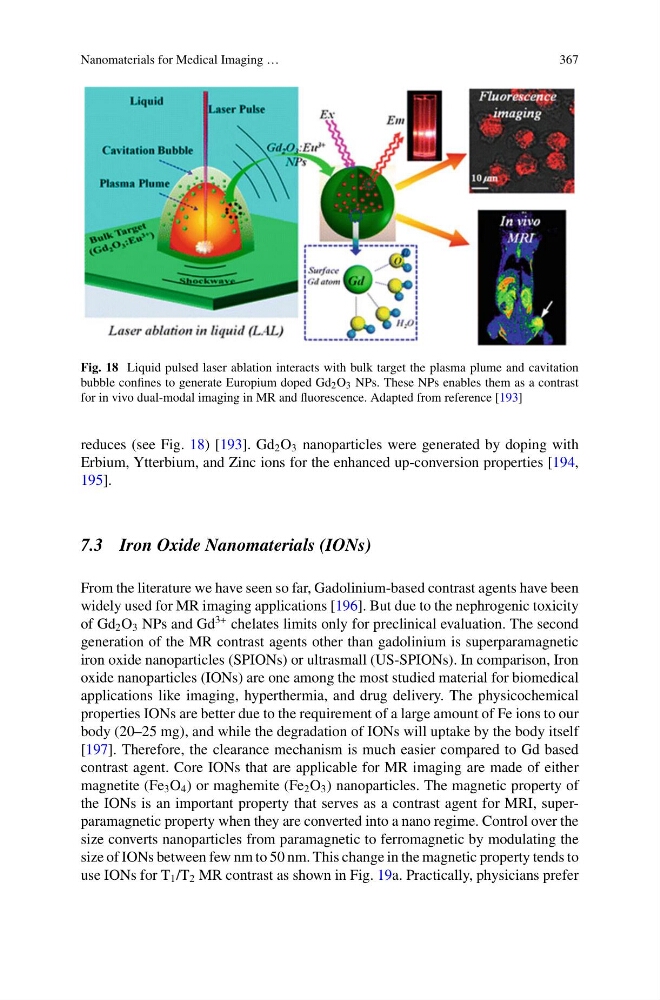
Fig. 18 Liquid pulsed laser ablation interacts with bulk target the plasma plume and cavitation
bubble confines to generate Europium doped Gd 2 O 3 NPs. These NPs enables them as a contrast
for in vivo dual-modal imaging in MR and fluorescence. Adapted from reference [193]
reduces (see Fig. 18) [193]. Gd 2 O 3 nanoparticles were generated by doping with
Erbium, Ytterbium, and Zinc ions for the enhanced up-conversion properties [194,
195].
7.3 Iron Oxide Nanomaterials (IONs)
From the literature we have seen so far, Gadolinium-based contrast agents have been
widely used for MR imaging applications [196]. But due to the nephrogenic toxicity
of Gd 2 O 3 NPs and Gd
3+ chelates limits only for preclinical evaluation. The second
generation of the MR contrast agents other than gadolinium is superparamagnetic
iron oxide nanoparticles (SPIONs) or ultrasmall (US-SPIONs). In comparison, Iron
oxide nanoparticles (IONs) are one among the most studied material for biomedical
applications like imaging, hyperthermia, and drug delivery. The physicochemical
properties IONs are better due to the requirement of a large amount of Fe ions to our
body (20–25 mg), and while the degradation of IONs will uptake by the body itself
[197]. Therefore, the clearance mechanism is much easier compared to Gd based
contrast agent. Core IONs that are applicable for MR imaging are made of either
magnetite (Fe 3 O 4 ) or maghemite (Fe 2 O 3 ) nanoparticles. The magnetic property of
the IONs is an important property that serves as a contrast agent for MRI, superparamagnetic property when they are converted into a nano regime. Control over the
size converts nanoparticles from paramagnetic to ferromagnetic by modulating the
size of IONs between few nm to 50 nm. This change in the magnetic property tends to
use IONs for T 1 /T 2 MR contrast as shown in Fig. 19a. Practically, physicians prefer
367
Fig. 18 Liquid pulsed laser ablation interacts with bulk target the plasma plume and cavitation
bubble confines to generate Europium doped Gd 2 O 3 NPs. These NPs enables them as a contrast
for in vivo dual-modal imaging in MR and fluorescence. Adapted from reference [193]
reduces (see Fig. 18) [193]. Gd 2 O 3 nanoparticles were generated by doping with
Erbium, Ytterbium, and Zinc ions for the enhanced up-conversion properties [194,
195].
7.3 Iron Oxide Nanomaterials (IONs)
From the literature we have seen so far, Gadolinium-based contrast agents have been
widely used for MR imaging applications [196]. But due to the nephrogenic toxicity
of Gd 2 O 3 NPs and Gd
3+ chelates limits only for preclinical evaluation. The second
generation of the MR contrast agents other than gadolinium is superparamagnetic
iron oxide nanoparticles (SPIONs) or ultrasmall (US-SPIONs). In comparison, Iron
oxide nanoparticles (IONs) are one among the most studied material for biomedical
applications like imaging, hyperthermia, and drug delivery. The physicochemical
properties IONs are better due to the requirement of a large amount of Fe ions to our
body (20–25 mg), and while the degradation of IONs will uptake by the body itself
[197]. Therefore, the clearance mechanism is much easier compared to Gd based
contrast agent. Core IONs that are applicable for MR imaging are made of either
magnetite (Fe 3 O 4 ) or maghemite (Fe 2 O 3 ) nanoparticles. The magnetic property of
the IONs is an important property that serves as a contrast agent for MRI, superparamagnetic property when they are converted into a nano regime. Control over the
size converts nanoparticles from paramagnetic to ferromagnetic by modulating the
size of IONs between few nm to 50 nm. This change in the magnetic property tends to
use IONs for T 1 /T 2 MR contrast as shown in Fig. 19a. Practically, physicians prefer