

Nanomaterials for Medical Imaging …
355
instance, the efficiency of Ta is 1.4 times higher than Iodine [110]. Both metallic and
oxide form of Ta was used as contrast agents as well in dentistry, orthopedics, and
regenerative medicine [111]. Tantalum is still considered as CT agent compared to
gold, which has similar X-ray attenuation properties like Bismuth. In 2012, Marino’s
group developed Zwitterionic polymer-coated Tantalum oxide (TaO) nanoparticles
as an X-ray contrast agent. Administering these particles at a dose of 1500 mg Ta kg
−1
had no significant impact on rodent physiology [112]. They were able to hypothesize
that the particles with size 2–3 nm would significantly accumulate in kidneys and
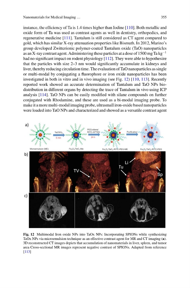
liver, thereby reducing circulation time. The evaluation of TaO nanoparticles as single
or multi-modal by conjugating a fluorophore or iron oxide nanoparticles has been
investigated in both in vitro and in vivo imaging (see Fig. 12) [110, 113]. Recently
reported work showed an accurate determination of Tantalum and TaO NPs biodistribution in different organs by detecting the trace of Tantalum in vivo using ICP
analysis [114]. TaO NPs can be easily modified with silane compounds on further
conjugated with Rhodamine, and these are used as a bi-modal imaging probe. To
make it a more multi-modal imaging probe, ultrasmall iron-oxide based nanoparticles
were loaded into TaO NPs and characterized and showed as a versatile contrast agent
Fig. 12 Multimodal Iron oxide NPs into TaOx NPs: Incorporating SPIONs while synthesizing
TaOx NPs via microemulsion technique as an effective contrast agent for MR and CT imaging (a).
3D reconstructed CT images depicts that accumulation of nanomaterials in liver, spleen, and tumor
area Cross-sectional MR images represent negative contrast of SPIONs. Adapted from reference
[113]
355
instance, the efficiency of Ta is 1.4 times higher than Iodine [110]. Both metallic and
oxide form of Ta was used as contrast agents as well in dentistry, orthopedics, and
regenerative medicine [111]. Tantalum is still considered as CT agent compared to
gold, which has similar X-ray attenuation properties like Bismuth. In 2012, Marino’s
group developed Zwitterionic polymer-coated Tantalum oxide (TaO) nanoparticles
as an X-ray contrast agent. Administering these particles at a dose of 1500 mg Ta kg
−1
had no significant impact on rodent physiology [112]. They were able to hypothesize
that the particles with size 2–3 nm would significantly accumulate in kidneys and
liver, thereby reducing circulation time. The evaluation of TaO nanoparticles as single
or multi-modal by conjugating a fluorophore or iron oxide nanoparticles has been
investigated in both in vitro and in vivo imaging (see Fig. 12) [110, 113]. Recently
reported work showed an accurate determination of Tantalum and TaO NPs biodistribution in different organs by detecting the trace of Tantalum in vivo using ICP
analysis [114]. TaO NPs can be easily modified with silane compounds on further
conjugated with Rhodamine, and these are used as a bi-modal imaging probe. To
make it a more multi-modal imaging probe, ultrasmall iron-oxide based nanoparticles
were loaded into TaO NPs and characterized and showed as a versatile contrast agent
Fig. 12 Multimodal Iron oxide NPs into TaOx NPs: Incorporating SPIONs while synthesizing
TaOx NPs via microemulsion technique as an effective contrast agent for MR and CT imaging (a).
3D reconstructed CT images depicts that accumulation of nanomaterials in liver, spleen, and tumor
area Cross-sectional MR images represent negative contrast of SPIONs. Adapted from reference
[113]