

340
N. Ashwin Kumar et al.
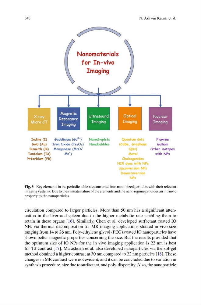
Fig. 3 Key elements in the periodic table are converted into nano-sized particles with their relevant
imaging systems. Due to their innate nature of the elements and the nano regime provides an intrinsic
property to the nanoparticles
circulation compared to larger particles. More than 50 nm has a significant attenuation in the liver and spleen due to the higher metabolic rate enabling them to
retain in these organs [16]. Similarly, Chen et al. developed surfactant coated IO
NPs via thermal decomposition for MR imaging applications studied in vivo size
ranging from 14 to 26 nm. Poly-ethylene glycol (PEG) coated IO nanoparticles have
shown better magnetic properties concerning the size. But the results provided that
the optimum size of IO NPs for the in vivo imaging application is 22 nm is best
for T2 contrast [17]. Marashdeh et al. also developed nanoparticles via the sol-gel
method obtained a higher contrast at 30 nm compared to 22 nm particles [18]. These
changes in MR contrast were not evident, and it can be concluded due to variation in
synthesis procedure, size due to surfactant, and poly-dispersity. Also, the nanoparticle
N. Ashwin Kumar et al.
Fig. 3 Key elements in the periodic table are converted into nano-sized particles with their relevant
imaging systems. Due to their innate nature of the elements and the nano regime provides an intrinsic
property to the nanoparticles
circulation compared to larger particles. More than 50 nm has a significant attenuation in the liver and spleen due to the higher metabolic rate enabling them to
retain in these organs [16]. Similarly, Chen et al. developed surfactant coated IO
NPs via thermal decomposition for MR imaging applications studied in vivo size
ranging from 14 to 26 nm. Poly-ethylene glycol (PEG) coated IO nanoparticles have
shown better magnetic properties concerning the size. But the results provided that
the optimum size of IO NPs for the in vivo imaging application is 22 nm is best
for T2 contrast [17]. Marashdeh et al. also developed nanoparticles via the sol-gel
method obtained a higher contrast at 30 nm compared to 22 nm particles [18]. These
changes in MR contrast were not evident, and it can be concluded due to variation in
synthesis procedure, size due to surfactant, and poly-dispersity. Also, the nanoparticle