

120
P. Hameed et al.
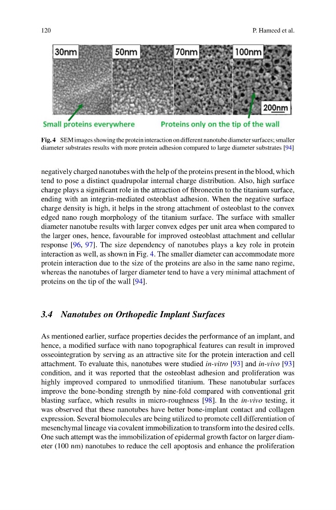
Fig. 4 SEM images showing the protein interaction on different nanotube diameter surfaces; smaller
diameter substrates results with more protein adhesion compared to large diameter substrates [94]
negatively charged nanotubes with the help of the proteins present in the blood, which
tend to pose a distinct quadrupolar internal charge distribution. Also, high surface
charge plays a significant role in the attraction of fibronectin to the titanium surface,
ending with an integrin-mediated osteoblast adhesion. When the negative surface
charge density is high, it helps in the strong attachment of osteoblast to the convex
edged nano rough morphology of the titanium surface. The surface with smaller
diameter nanotube results with larger convex edges per unit area when compared to
the larger ones, hence, favourable for improved osteoblast attachment and cellular
response [96, 97]. The size dependency of nanotubes plays a key role in protein
interaction as well, as shown in Fig. 4. The smaller diameter can accommodate more
protein interaction due to the size of the proteins are also in the same nano regime,
whereas the nanotubes of larger diameter tend to have a very minimal attachment of
proteins on the tip of the wall [94].
3.4 Nanotubes on Orthopedic Implant Surfaces
As mentioned earlier, surface properties decides the performance of an implant, and
hence, a modified surface with nano topographical features can result in improved
osseointegration by serving as an attractive site for the protein interaction and cell
attachment. To evaluate this, nanotubes were studied in-vitro [93] and in-vivo [93]
condition, and it was reported that the osteoblast adhesion and proliferation was
highly improved compared to unmodified titanium. These nanotubular surfaces
improve the bone-bonding strength by nine-fold compared with conventional grit
blasting surface, which results in micro-roughness [98]. In the in-vivo testing, it
was observed that these nanotubes have better bone-implant contact and collagen
expression. Several biomolecules are being utilized to promote cell differentiation of
mesenchymal lineage via covalent immobilization to transform into the desired cells.
One such attempt was the immobilization of epidermal growth factor on larger diameter (100 nm) nanotubes to reduce the cell apoptosis and enhance the proliferation
P. Hameed et al.
Fig. 4 SEM images showing the protein interaction on different nanotube diameter surfaces; smaller
diameter substrates results with more protein adhesion compared to large diameter substrates [94]
negatively charged nanotubes with the help of the proteins present in the blood, which
tend to pose a distinct quadrupolar internal charge distribution. Also, high surface
charge plays a significant role in the attraction of fibronectin to the titanium surface,
ending with an integrin-mediated osteoblast adhesion. When the negative surface
charge density is high, it helps in the strong attachment of osteoblast to the convex
edged nano rough morphology of the titanium surface. The surface with smaller
diameter nanotube results with larger convex edges per unit area when compared to
the larger ones, hence, favourable for improved osteoblast attachment and cellular
response [96, 97]. The size dependency of nanotubes plays a key role in protein
interaction as well, as shown in Fig. 4. The smaller diameter can accommodate more
protein interaction due to the size of the proteins are also in the same nano regime,
whereas the nanotubes of larger diameter tend to have a very minimal attachment of
proteins on the tip of the wall [94].
3.4 Nanotubes on Orthopedic Implant Surfaces
As mentioned earlier, surface properties decides the performance of an implant, and
hence, a modified surface with nano topographical features can result in improved
osseointegration by serving as an attractive site for the protein interaction and cell
attachment. To evaluate this, nanotubes were studied in-vitro [93] and in-vivo [93]
condition, and it was reported that the osteoblast adhesion and proliferation was
highly improved compared to unmodified titanium. These nanotubular surfaces
improve the bone-bonding strength by nine-fold compared with conventional grit
blasting surface, which results in micro-roughness [98]. In the in-vivo testing, it
was observed that these nanotubes have better bone-implant contact and collagen
expression. Several biomolecules are being utilized to promote cell differentiation of
mesenchymal lineage via covalent immobilization to transform into the desired cells.
One such attempt was the immobilization of epidermal growth factor on larger diameter (100 nm) nanotubes to reduce the cell apoptosis and enhance the proliferation