

Nanofibers and Nanosurfaces
119
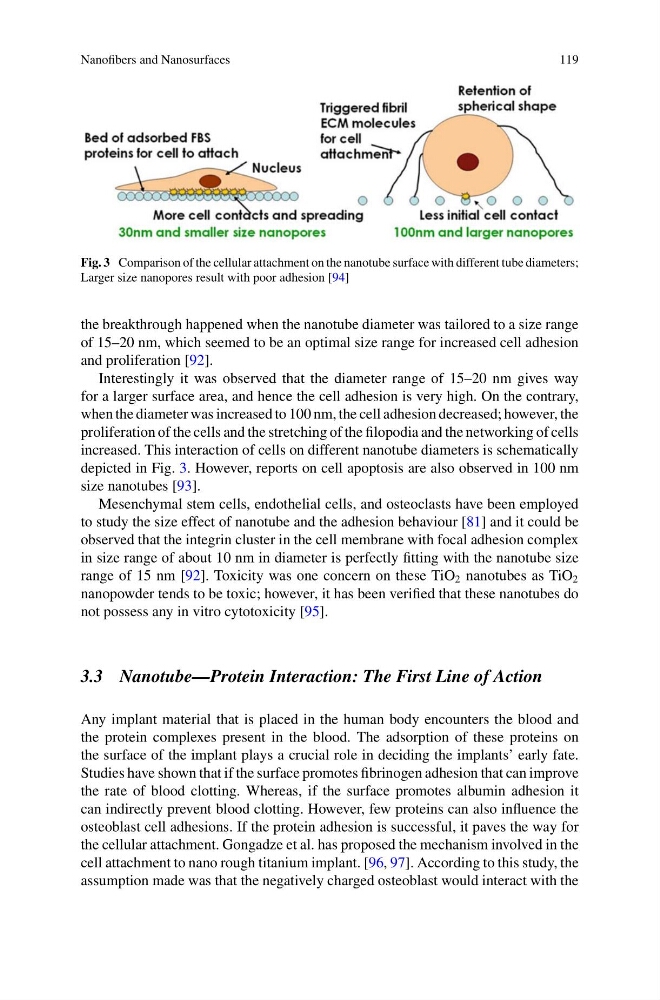
Fig. 3 Comparison of the cellular attachment on the nanotube surface with different tube diameters;
Larger size nanopores result with poor adhesion [94]
the breakthrough happened when the nanotube diameter was tailored to a size range
of 15–20 nm, which seemed to be an optimal size range for increased cell adhesion
and proliferation [92].
Interestingly it was observed that the diameter range of 15–20 nm gives way
for a larger surface area, and hence the cell adhesion is very high. On the contrary,
when the diameter was increased to 100 nm, the cell adhesion decreased; however, the
proliferation of the cells and the stretching of the filopodia and the networking of cells
increased. This interaction of cells on different nanotube diameters is schematically
depicted in Fig. 3. However, reports on cell apoptosis are also observed in 100 nm
size nanotubes [93].
Mesenchymal stem cells, endothelial cells, and osteoclasts have been employed
to study the size effect of nanotube and the adhesion behaviour [81] and it could be
observed that the integrin cluster in the cell membrane with focal adhesion complex
in size range of about 10 nm in diameter is perfectly fitting with the nanotube size
range of 15 nm [92]. Toxicity was one concern on these TiO 2 nanotubes as TiO 2
nanopowder tends to be toxic; however, it has been verified that these nanotubes do
not possess any in vitro cytotoxicity [95].
3.3 Nanotube—Protein Interaction: The First Line of Action
Any implant material that is placed in the human body encounters the blood and
the protein complexes present in the blood. The adsorption of these proteins on
the surface of the implant plays a crucial role in deciding the implants’ early fate.
Studies have shown that if the surface promotes fibrinogen adhesion that can improve
the rate of blood clotting. Whereas, if the surface promotes albumin adhesion it
can indirectly prevent blood clotting. However, few proteins can also influence the
osteoblast cell adhesions. If the protein adhesion is successful, it paves the way for
the cellular attachment. Gongadze et al. has proposed the mechanism involved in the
cell attachment to nano rough titanium implant. [96, 97]. According to this study, the
assumption made was that the negatively charged osteoblast would interact with the
119
Fig. 3 Comparison of the cellular attachment on the nanotube surface with different tube diameters;
Larger size nanopores result with poor adhesion [94]
the breakthrough happened when the nanotube diameter was tailored to a size range
of 15–20 nm, which seemed to be an optimal size range for increased cell adhesion
and proliferation [92].
Interestingly it was observed that the diameter range of 15–20 nm gives way
for a larger surface area, and hence the cell adhesion is very high. On the contrary,
when the diameter was increased to 100 nm, the cell adhesion decreased; however, the
proliferation of the cells and the stretching of the filopodia and the networking of cells
increased. This interaction of cells on different nanotube diameters is schematically
depicted in Fig. 3. However, reports on cell apoptosis are also observed in 100 nm
size nanotubes [93].
Mesenchymal stem cells, endothelial cells, and osteoclasts have been employed
to study the size effect of nanotube and the adhesion behaviour [81] and it could be
observed that the integrin cluster in the cell membrane with focal adhesion complex
in size range of about 10 nm in diameter is perfectly fitting with the nanotube size
range of 15 nm [92]. Toxicity was one concern on these TiO 2 nanotubes as TiO 2
nanopowder tends to be toxic; however, it has been verified that these nanotubes do
not possess any in vitro cytotoxicity [95].
3.3 Nanotube—Protein Interaction: The First Line of Action
Any implant material that is placed in the human body encounters the blood and
the protein complexes present in the blood. The adsorption of these proteins on
the surface of the implant plays a crucial role in deciding the implants’ early fate.
Studies have shown that if the surface promotes fibrinogen adhesion that can improve
the rate of blood clotting. Whereas, if the surface promotes albumin adhesion it
can indirectly prevent blood clotting. However, few proteins can also influence the
osteoblast cell adhesions. If the protein adhesion is successful, it paves the way for
the cellular attachment. Gongadze et al. has proposed the mechanism involved in the
cell attachment to nano rough titanium implant. [96, 97]. According to this study, the
assumption made was that the negatively charged osteoblast would interact with the