

650
S. P. Singh and S. Siddhanta
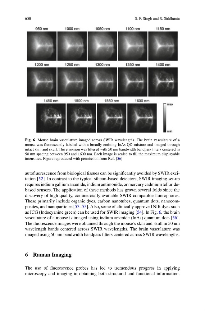
Fig. 6 Mouse brain vasculature imaged across SWIR wavelengths. The brain vasculature of a
mouse was fluorescently labeled with a broadly emitting InAs QD mixture and imaged through
intact skin and skull. The emission was filtered with 50 nm bandwidth bandpass filters centered in
50 nm spacing between 950 and 1600 nm. Each image is scaled to fill the maximum displayable
intensities. Figure reproduced with permission from Ref. [56]
autofluorescence from biological tissues can be significantly avoided by SWIR excitation [52]. In contrast to the typical silicon-based detectors, SWIR imaging set-up
requires indium gallium arsenide, indium antimonide, or mercury cadmium telluridebased sensors. The application of these methods has grown several folds since the
discovery of high quality, commercially available SWIR compatible fluorophores.
These primarily include organic dyes, carbon nanotubes, quantum dots, nanocomposites, and nanoparticles [53–55]. Also, some of clinically approved NIR dyes such
as ICG (Indocyanine green) can be used for SWIR imaging [54]. In Fig. 6, the brain
vasculature of a mouse is imaged using indium arsenide (InAs) quantum dots [56].
The fluorescence images were obtained through the mouse’s skin and skull in 50 nm
wavelength bands centered across SWIR wavelengths. The brain vasculature was
imaged using 50 nm bandwidth bandpass filters centered across SWIR wavelengths.
6 Raman Imaging
The use of fluorescence probes has led to tremendous progress in applying
microscopy and imaging in obtaining both structural and functional information.