

Optical Imaging in Biology: Basics and Applications
647
following a naked fluorophore to probe the cellular environment. The first application of FCS was demonstrated in studying drug intercalation in nucleic acids, later
it was applied to different streams of biological sciences [32]. Some of the common
applications include monitoring protein, lipid diffusion of across the cell membrane,
following binding efficiency of surface receptors and molecules, studying proteinprotein or protein-nucleic acid, lipid-protein interactions, intracellular movement of
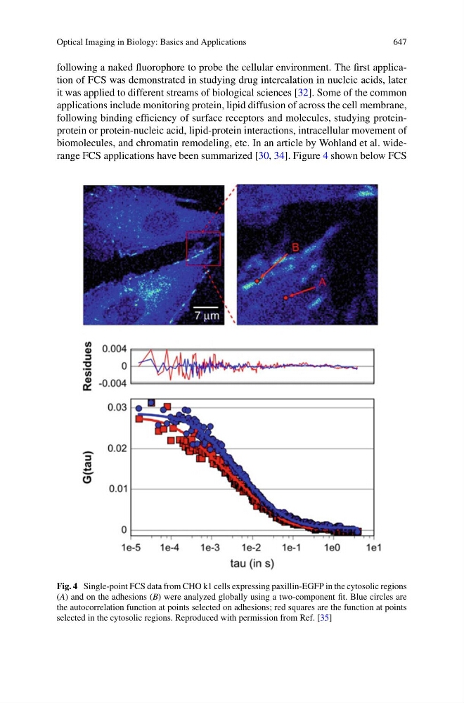
biomolecules, and chromatin remodeling, etc. In an article by Wohland et al. widerange FCS applications have been summarized [30, 34]. Figure 4 shown below FCS
Fig. 4 Single-point FCS data from CHO k1 cells expressing paxillin-EGFP in the cytosolic regions
(A) and on the adhesions (B) were analyzed globally using a two-component fit. Blue circles are
the autocorrelation function at points selected on adhesions; red squares are the function at points
selected in the cytosolic regions. Reproduced with permission from Ref. [35]