

586
J. Jaisawal et al.
by AFM in different modes like contact, tapping and non-contact mode which has
resolution in the nanoscale. AFM is one of most surface sensitive technique that
can be used to generate high resolution images. Basic principle of this technique
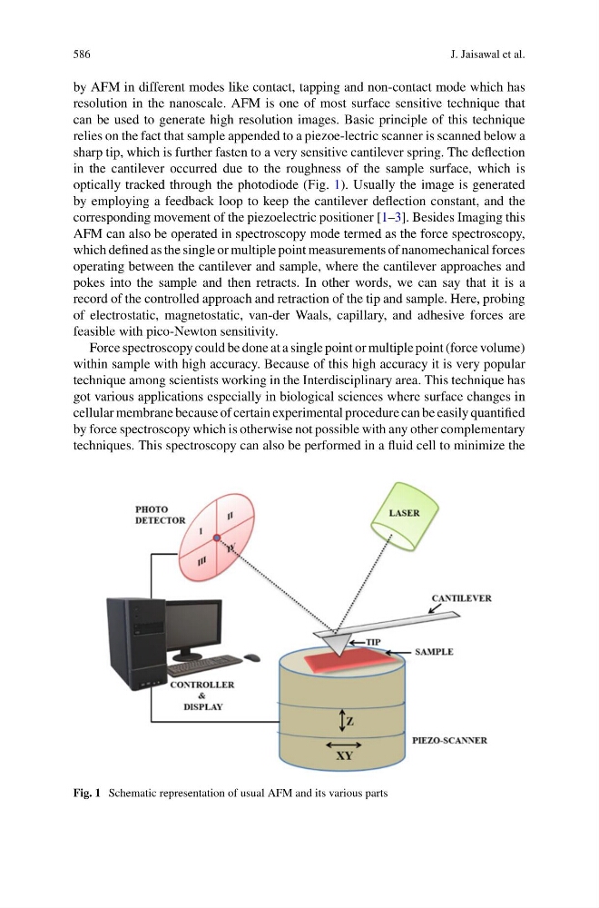
relies on the fact that sample appended to a piezoe-lectric scanner is scanned below a
sharp tip, which is further fasten to a very sensitive cantilever spring. The deflection
in the cantilever occurred due to the roughness of the sample surface, which is
optically tracked through the photodiode (Fig. 1). Usually the image is generated
by employing a feedback loop to keep the cantilever deflection constant, and the
corresponding movement of the piezoelectric positioner [1–3]. Besides Imaging this
AFM can also be operated in spectroscopy mode termed as the force spectroscopy,
which defined as the single or multiple point measurements of nanomechanical forces
operating between the cantilever and sample, where the cantilever approaches and
pokes into the sample and then retracts. In other words, we can say that it is a
record of the controlled approach and retraction of the tip and sample. Here, probing
of electrostatic, magnetostatic, van-der Waals, capillary, and adhesive forces are
feasible with pico-Newton sensitivity.
Force spectroscopy could be done at a single point or multiple point (force volume)
within sample with high accuracy. Because of this high accuracy it is very popular
technique among scientists working in the Interdisciplinary area. This technique has
got various applications especially in biological sciences where surface changes in
cellular membrane because of certain experimental procedure can be easily quantified
by force spectroscopy which is otherwise not possible with any other complementary
techniques. This spectroscopy can also be performed in a fluid cell to minimize the
Fig. 1 Schematic representation of usual AFM and its various parts
J. Jaisawal et al.
by AFM in different modes like contact, tapping and non-contact mode which has
resolution in the nanoscale. AFM is one of most surface sensitive technique that
can be used to generate high resolution images. Basic principle of this technique
relies on the fact that sample appended to a piezoe-lectric scanner is scanned below a
sharp tip, which is further fasten to a very sensitive cantilever spring. The deflection
in the cantilever occurred due to the roughness of the sample surface, which is
optically tracked through the photodiode (Fig. 1). Usually the image is generated
by employing a feedback loop to keep the cantilever deflection constant, and the
corresponding movement of the piezoelectric positioner [1–3]. Besides Imaging this
AFM can also be operated in spectroscopy mode termed as the force spectroscopy,
which defined as the single or multiple point measurements of nanomechanical forces
operating between the cantilever and sample, where the cantilever approaches and
pokes into the sample and then retracts. In other words, we can say that it is a
record of the controlled approach and retraction of the tip and sample. Here, probing
of electrostatic, magnetostatic, van-der Waals, capillary, and adhesive forces are
feasible with pico-Newton sensitivity.
Force spectroscopy could be done at a single point or multiple point (force volume)
within sample with high accuracy. Because of this high accuracy it is very popular
technique among scientists working in the Interdisciplinary area. This technique has
got various applications especially in biological sciences where surface changes in
cellular membrane because of certain experimental procedure can be easily quantified
by force spectroscopy which is otherwise not possible with any other complementary
techniques. This spectroscopy can also be performed in a fluid cell to minimize the
Fig. 1 Schematic representation of usual AFM and its various parts