

Fundamentals of ATR-FTIR Spectroscopy and Its Role …
27
DMF molecules tend to cluster among themselves at higher concentrations at the
crystal surface projecting significant spectral changes.
4.4 Application in Forensic Sciences
Over the course of a few years, ATR-FTIR spectroscopy has acquired the potency in
differentiating the constituents and estimating their degradative states at the molecular level. It has been therefore applied not only for the biomolecular probing but
also in the domain of forensic chemometrics, identifying toxic chemicals, and the
bio-threat or warfare agents for forensic analysis [71–74]. Considering the simplified
mode of working of the ATR-FTIR technique, it is now being widely commercialized
as a hand-held portable testing tool for on-site evaluation purposes especially in the
field of forensic sciences [75]. Many reports reveal its importance in quantitative and
trace-level identification of elements with a non-destructive approach which include
discriminating paper elements and its age, drug profiling and quantitation, probing
sunscreen stains, predicting the sex of an individual through fingernail analyses, etc.
[71, 72, 76–78].
Zou et al. [79] unraveled the potential of the ATR-FTIR spectroscopic tool along
with the Raman spectroscopy to identify and differentiate between the human blood
and semen sample as well as their deterioration state. Spotting of the biofluid presence and their preservation is pivotal for further examination of the crime scene
conditions. In this work, the authors utilized a single reflection ATR-FTIR spectrometric geometry with a horizontal ZnSe crystal mount accessory. The sample slides
containing the body fluid sample were attached to the crystal surface via clamping.
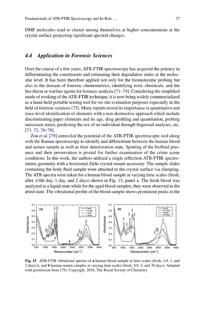
The ATR spectra were taken for a human blood sample at varying time scales (fresh,
after 1/4th day, 1 day, and 2 days) shown in Fig. 15, panel a. The fresh blood was
analyzed in a liquid state while for the aged blood samples, they were observed in the
dried state. The vibrational profile of the blood sample shows prominent peaks in the
Fig. 15 ATR-FTIR vibrational spectra of a human blood sample at time scales (fresh, 1/4, 1, and
2 day(s)), and b human semen samples at varying time scales (fresh, 5/4, 5, and 30 days). Adapted
with permission from [79]. Copyright, 2016, The Royal Society of Chemistry
27
DMF molecules tend to cluster among themselves at higher concentrations at the
crystal surface projecting significant spectral changes.
4.4 Application in Forensic Sciences
Over the course of a few years, ATR-FTIR spectroscopy has acquired the potency in
differentiating the constituents and estimating their degradative states at the molecular level. It has been therefore applied not only for the biomolecular probing but
also in the domain of forensic chemometrics, identifying toxic chemicals, and the
bio-threat or warfare agents for forensic analysis [71–74]. Considering the simplified
mode of working of the ATR-FTIR technique, it is now being widely commercialized
as a hand-held portable testing tool for on-site evaluation purposes especially in the
field of forensic sciences [75]. Many reports reveal its importance in quantitative and
trace-level identification of elements with a non-destructive approach which include
discriminating paper elements and its age, drug profiling and quantitation, probing
sunscreen stains, predicting the sex of an individual through fingernail analyses, etc.
[71, 72, 76–78].
Zou et al. [79] unraveled the potential of the ATR-FTIR spectroscopic tool along
with the Raman spectroscopy to identify and differentiate between the human blood
and semen sample as well as their deterioration state. Spotting of the biofluid presence and their preservation is pivotal for further examination of the crime scene
conditions. In this work, the authors utilized a single reflection ATR-FTIR spectrometric geometry with a horizontal ZnSe crystal mount accessory. The sample slides
containing the body fluid sample were attached to the crystal surface via clamping.
The ATR spectra were taken for a human blood sample at varying time scales (fresh,
after 1/4th day, 1 day, and 2 days) shown in Fig. 15, panel a. The fresh blood was
analyzed in a liquid state while for the aged blood samples, they were observed in the
dried state. The vibrational profile of the blood sample shows prominent peaks in the
Fig. 15 ATR-FTIR vibrational spectra of a human blood sample at time scales (fresh, 1/4, 1, and
2 day(s)), and b human semen samples at varying time scales (fresh, 5/4, 5, and 30 days). Adapted
with permission from [79]. Copyright, 2016, The Royal Society of Chemistry