

Photon Upconversion Spectroscopy
403
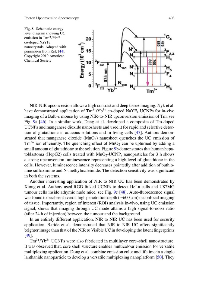
Fig. 8 Schematic energy
level diagram showing UC
emission in Tm 3+ /Yb 3+
co-doped NaYF 4
nanocrystals. Adapted with
permission from Ref. [44].
Copyright 2010 American
Chemical Society
NIR-NIR upconversion allows a high contrast and deep tissue imaging. Nyk et al.
have demonstrated application of Tm
3+ /Yb
3+ co-doped NaYF 4 UCNPs for in-vivo
imaging of a Balb-c mouse by using NIR-to-NIR upconversion emission of Tm, see
Fig. 9a [46]. In a similar work, Deng et al. developed a composite of Tm-doped
UCNPs and manganese dioxide nanosheets and used it for rapid and selective detection of glutathione in aqueous solutions and in living cells [47]. Authors demonstrated that manganese dioxide (MnO 2 ) nanosheet quenches the UC emission of
Tm
3+ ion efficiently. The quenching effect of MnO 2 can be upturned by adding a
small amount of glutathione to the solution. Figure 9b demonstrates that human hepatoblastoma (HepG2) cells treated with MnO 2 -UCNP s nanoparticles for 3 h shows
a strong upconversion luminescence representing a high level of glutathione in the
cells. However, luminescence intensity decreases pointedly after addition of buthionine sulfoximine and N-methylmaleimide. The detection sensitivity was significant
in both the systems.
Another interesting application of NIR to NIR UC has been demonstrated by
Xiong et al. Authors used RGD linked UCNPs to detect HeLa cells and U87MG
tumour cells inside athymic nude mice, see Fig. 9c [48]. Auto-fluorescence signal
was found to be absent even at high penetration depth (∼600 μm) in confocal imaging
of tissue. Importantly, region of interest (ROI) analysis in-vivo, using UC emission
signal, shows that imaging through UC mode attains a high signal-to-noise ratio
(after 24 h of injection) between the tumour and the background.
In an entirely different application, NIR to NIR UC has been used for security
application. Baride et al. demonstrated that NIR to NIR UC offers significantly
brighter image than that of the NIR to Visible UC in developing the latent fingerprints
[49].
Tm
3+ /Yb
3+ UCNPs were also fabricated in multilayer core–shell nanostructure.
It was observed that, core shell structure enables multicolour emission for versatile
multiplexing application. Dong et al. combine emission color and lifetime in a single
lanthanide nanoparticle to develop a versatile multiplexing nanoplatform [50]. They
403
Fig. 8 Schematic energy
level diagram showing UC
emission in Tm 3+ /Yb 3+
co-doped NaYF 4
nanocrystals. Adapted with
permission from Ref. [44].
Copyright 2010 American
Chemical Society
NIR-NIR upconversion allows a high contrast and deep tissue imaging. Nyk et al.
have demonstrated application of Tm
3+ /Yb
3+ co-doped NaYF 4 UCNPs for in-vivo
imaging of a Balb-c mouse by using NIR-to-NIR upconversion emission of Tm, see
Fig. 9a [46]. In a similar work, Deng et al. developed a composite of Tm-doped
UCNPs and manganese dioxide nanosheets and used it for rapid and selective detection of glutathione in aqueous solutions and in living cells [47]. Authors demonstrated that manganese dioxide (MnO 2 ) nanosheet quenches the UC emission of
Tm
3+ ion efficiently. The quenching effect of MnO 2 can be upturned by adding a
small amount of glutathione to the solution. Figure 9b demonstrates that human hepatoblastoma (HepG2) cells treated with MnO 2 -UCNP s nanoparticles for 3 h shows
a strong upconversion luminescence representing a high level of glutathione in the
cells. However, luminescence intensity decreases pointedly after addition of buthionine sulfoximine and N-methylmaleimide. The detection sensitivity was significant
in both the systems.
Another interesting application of NIR to NIR UC has been demonstrated by
Xiong et al. Authors used RGD linked UCNPs to detect HeLa cells and U87MG
tumour cells inside athymic nude mice, see Fig. 9c [48]. Auto-fluorescence signal
was found to be absent even at high penetration depth (∼600 μm) in confocal imaging
of tissue. Importantly, region of interest (ROI) analysis in-vivo, using UC emission
signal, shows that imaging through UC mode attains a high signal-to-noise ratio
(after 24 h of injection) between the tumour and the background.
In an entirely different application, NIR to NIR UC has been used for security
application. Baride et al. demonstrated that NIR to NIR UC offers significantly
brighter image than that of the NIR to Visible UC in developing the latent fingerprints
[49].
Tm
3+ /Yb
3+ UCNPs were also fabricated in multilayer core–shell nanostructure.
It was observed that, core shell structure enables multicolour emission for versatile
multiplexing application. Dong et al. combine emission color and lifetime in a single
lanthanide nanoparticle to develop a versatile multiplexing nanoplatform [50]. They