

Fundamentals and Applications of Surface Enhanced Raman …
199
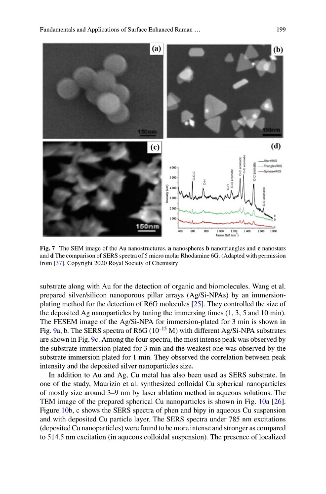
Fig. 7 The SEM image of the Au nanostructures. a nanospheres b nanotriangles and c nanostars
and d The comparison of SERS spectra of 5 micro molar Rhodamine 6G. (Adapted with permission
from [37]. Copyright 2020 Royal Society of Chemistry
substrate along with Au for the detection of organic and biomolecules. Wang et al.
prepared silver/silicon nanoporous pillar arrays (Ag/Si-NPAs) by an immersionplating method for the detection of R6G molecules [25]. They controlled the size of
the deposited Ag nanoparticles by tuning the immersing times (1, 3, 5 and 10 min).
The FESEM image of the Ag/Si-NPA for immersion-plated for 3 min is shown in
Fig. 9a, b. The SERS spectra of R6G (10
–15 M) with different Ag/Si-NPA substrates
are shown in Fig. 9c. Among the four spectra, the most intense peak was observed by
the substrate immersion plated for 3 min and the weakest one was observed by the
substrate immersion plated for 1 min. They observed the correlation between peak
intensity and the deposited silver nanoparticles size.
In addition to Au and Ag, Cu metal has also been used as SERS substrate. In
one of the study, Maurizio et al. synthesized colloidal Cu spherical nanoparticles
of mostly size around 3–9 nm by laser ablation method in aqueous solutions. The
TEM image of the prepared spherical Cu nanoparticles is shown in Fig. 10a [26].
Figure 10b, c shows the SERS spectra of phen and bipy in aqueous Cu suspension
and with deposited Cu particle layer. The SERS spectra under 785 nm excitations
(deposited Cu nanoparticles) were found to be more intense and stronger as compared
to 514.5 nm excitation (in aqueous colloidal suspension). The presence of localized
199
Fig. 7 The SEM image of the Au nanostructures. a nanospheres b nanotriangles and c nanostars
and d The comparison of SERS spectra of 5 micro molar Rhodamine 6G. (Adapted with permission
from [37]. Copyright 2020 Royal Society of Chemistry
substrate along with Au for the detection of organic and biomolecules. Wang et al.
prepared silver/silicon nanoporous pillar arrays (Ag/Si-NPAs) by an immersionplating method for the detection of R6G molecules [25]. They controlled the size of
the deposited Ag nanoparticles by tuning the immersing times (1, 3, 5 and 10 min).
The FESEM image of the Ag/Si-NPA for immersion-plated for 3 min is shown in
Fig. 9a, b. The SERS spectra of R6G (10
–15 M) with different Ag/Si-NPA substrates
are shown in Fig. 9c. Among the four spectra, the most intense peak was observed by
the substrate immersion plated for 3 min and the weakest one was observed by the
substrate immersion plated for 1 min. They observed the correlation between peak
intensity and the deposited silver nanoparticles size.
In addition to Au and Ag, Cu metal has also been used as SERS substrate. In
one of the study, Maurizio et al. synthesized colloidal Cu spherical nanoparticles
of mostly size around 3–9 nm by laser ablation method in aqueous solutions. The
TEM image of the prepared spherical Cu nanoparticles is shown in Fig. 10a [26].
Figure 10b, c shows the SERS spectra of phen and bipy in aqueous Cu suspension
and with deposited Cu particle layer. The SERS spectra under 785 nm excitations
(deposited Cu nanoparticles) were found to be more intense and stronger as compared
to 514.5 nm excitation (in aqueous colloidal suspension). The presence of localized