

iron plaque has been reported from Pleistocene sediment in Aichi Prefecture, Japan
(Yoshida and Matsuoka 2004).
A polished section was used to examine the elemental distribution in the cylindrical iron mottles. Figure 5.7a shows a cross-sectional scanning electron microscope (SEM) image of a cylindrical iron mottle, which appears as a bright and thick
ring. The EDX spectra in Fig. 5.7b, c were obtained from the locations outlined in
Fig. 5.7a by dashed squares (b) (cylindrical iron mottle) and (c) (bulk soil outside the
iron mottle), respectively. The concentration of iron is much higher in the iron mottle
than in the outer soil matrix. In contrast, the concentrations of Al and Si are similar,
suggesting that the cylindrical iron mottle developed in the soil matrix directly
outside the plant root. Element maps of Si (Fig. 5.7d) and Fe (Fig. 5.7e) support
the interpretation of these EDX spectra. Although the Si concentration in the iron
mottle appears to be slightly lower than that of the bulk soil, the distribution pattern
of Si-rich particles in the iron mottle is the same as that of the bulk soil. The iron
concentration in the iron mottle is significantly higher than that of the bulk soil, and
the iron distribution pattern compares closely to the SEM image of the iron mottle
(Fig. 5.7a). Potentially, the EDX spectrum for the iron mottle (Fig. 5.7b) has very
small peaks of P and S that are lacking in the bulk soil EDX spectrum (Fig. 5.7c).
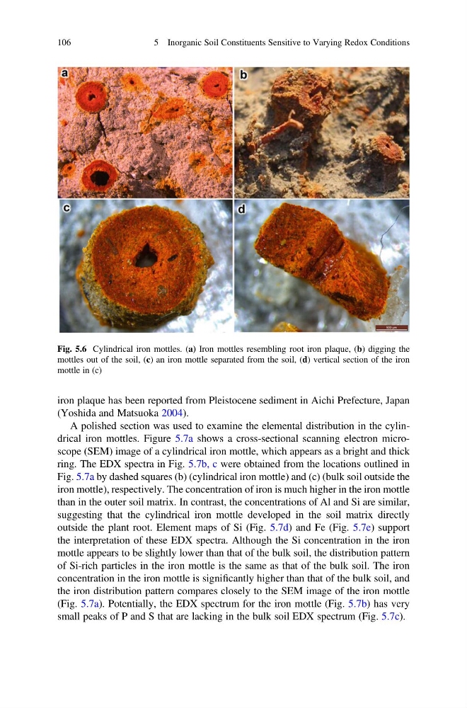
Fig. 5.6 Cylindrical iron mottles. (a) Iron mottles resembling root iron plaque, (b) digging the
mottles out of the soil, (c) an iron mottle separated from the soil, (d) vertical section of the iron
mottle in (c)
106
5 Inorganic Soil Constituents Sensitive to Varying Redox Conditions
(Yoshida and Matsuoka 2004).
A polished section was used to examine the elemental distribution in the cylindrical iron mottles. Figure 5.7a shows a cross-sectional scanning electron microscope (SEM) image of a cylindrical iron mottle, which appears as a bright and thick
ring. The EDX spectra in Fig. 5.7b, c were obtained from the locations outlined in
Fig. 5.7a by dashed squares (b) (cylindrical iron mottle) and (c) (bulk soil outside the
iron mottle), respectively. The concentration of iron is much higher in the iron mottle
than in the outer soil matrix. In contrast, the concentrations of Al and Si are similar,
suggesting that the cylindrical iron mottle developed in the soil matrix directly
outside the plant root. Element maps of Si (Fig. 5.7d) and Fe (Fig. 5.7e) support
the interpretation of these EDX spectra. Although the Si concentration in the iron
mottle appears to be slightly lower than that of the bulk soil, the distribution pattern
of Si-rich particles in the iron mottle is the same as that of the bulk soil. The iron
concentration in the iron mottle is significantly higher than that of the bulk soil, and
the iron distribution pattern compares closely to the SEM image of the iron mottle
(Fig. 5.7a). Potentially, the EDX spectrum for the iron mottle (Fig. 5.7b) has very
small peaks of P and S that are lacking in the bulk soil EDX spectrum (Fig. 5.7c).
Fig. 5.6 Cylindrical iron mottles. (a) Iron mottles resembling root iron plaque, (b) digging the
mottles out of the soil, (c) an iron mottle separated from the soil, (d) vertical section of the iron
mottle in (c)
106
5 Inorganic Soil Constituents Sensitive to Varying Redox Conditions