

PEPTIDE BIOSYNTHESIS
537
Cyclosporin A contains 11 amino acids, joined in a cyclic structure by peptide bonds. The structure is also
stabilized by intramolecular hydrogen bonds. Only two of the amino acids, i.e. alanine and valine, are typical of
proteins. The compound contains several N-methylated amino acid residues, together with the even less common
L-α-aminobutyric acid and an N-methylated butenylmethylthreonine. There is one D-amino acid, i.e. D-alanine, and
the assembly of the polypeptide chain is known to start from this residue. Many of the other natural cyclosporin
structures differ only with respect to a single amino acid (the α-aminobutyric acid residue) or the number of
amino acids that have the extra N-methyl group.
Of all the natural analogues, and many synthetic ones produced, cyclosporin A is the most valuable for drug use,
under the drug name ciclosporin. It is now widely exploited in organ and tissue transplant surgery, to prevent
rejection following bone marrow, kidney, liver, and heart transplants. It has revolutionized organ transplant
surgery, substantially increasing survival rates in transplant patients. It is believed to inhibit T-cell activation in
the immunosuppressive mechanism by first binding to a receptor protein, giving a complex that then inhibits a
phosphatase enzyme called calcineurin. The resultant aberrant phosphorylation reactions prevent appropriate gene
transcription and subsequent T-cell activation.
Box 13.11
Penicillins and cephalosporins are modified tripeptides
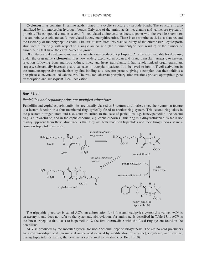
Penicillin and cephalosporin antibiotics are usually classed as β-lactam antibiotics, since their common feature
is a lactam function in a four-membered ring, typically fused to another ring system. This second ring takes in
the β-lactam nitrogen atom and also contains sulfur. In the case of penicillins, e.g. benzylpenicillin, the second
ring is a thiazolidine, and in the cephalosporins, e.g. cephalosporin C, this ring is a dihydrothiazine. What is not
readily apparent from these structures is that they are both modified tripeptides and their biosyntheses share a
common tripeptide precursor.
H 2 N
CO 2 H
O
H
N
NH
O
CO 2 H
SH
ACV
H 2 N
CO 2 H
O
H
N
N
O
S
CO 2 H
H
isopenicillin N
formation of fused
ring system
H
N
N
O
S
CO 2 H
H
O
benzylpenicillin
(penicillin G)
1
2
3
4
5
7
α-aminoadipic acid
6
acyltransferase
H 2 N
CO 2 H
O
H
N
N
O
H
S
CO 2 H
O
O
cephalosporin C
1
2
3
4
5
6
7
8
via ring expansion
process
L
L
D
PhCH 2 COSCoA
The tripeptide precursor is called ACV, an abbreviation for δ-(L-α-aminoadipyl)-L-cysteinyl-D-valine. ACV is
an acronym, and does not refer to the systematic abbreviations for amino acids described in Table 13.1. ACV is
the linear tripeptide that leads to isopenicillin N, the first intermediate with the fused-ring system found in the
penicillins.
ACV is produced by the modular system for non-ribosomal peptide biosynthesis. The amino acid precursors
are L-α-aminoadipic acid (an unusual amino acid derived by modification of L-lysine), L-cysteine, and L-valine;
during tripeptide formation, the L-valine is epimerized to D-valine (see Box 10.10).
537
Cyclosporin A contains 11 amino acids, joined in a cyclic structure by peptide bonds. The structure is also
stabilized by intramolecular hydrogen bonds. Only two of the amino acids, i.e. alanine and valine, are typical of
proteins. The compound contains several N-methylated amino acid residues, together with the even less common
L-α-aminobutyric acid and an N-methylated butenylmethylthreonine. There is one D-amino acid, i.e. D-alanine, and
the assembly of the polypeptide chain is known to start from this residue. Many of the other natural cyclosporin
structures differ only with respect to a single amino acid (the α-aminobutyric acid residue) or the number of
amino acids that have the extra N-methyl group.
Of all the natural analogues, and many synthetic ones produced, cyclosporin A is the most valuable for drug use,
under the drug name ciclosporin. It is now widely exploited in organ and tissue transplant surgery, to prevent
rejection following bone marrow, kidney, liver, and heart transplants. It has revolutionized organ transplant
surgery, substantially increasing survival rates in transplant patients. It is believed to inhibit T-cell activation in
the immunosuppressive mechanism by first binding to a receptor protein, giving a complex that then inhibits a
phosphatase enzyme called calcineurin. The resultant aberrant phosphorylation reactions prevent appropriate gene
transcription and subsequent T-cell activation.
Box 13.11
Penicillins and cephalosporins are modified tripeptides
Penicillin and cephalosporin antibiotics are usually classed as β-lactam antibiotics, since their common feature
is a lactam function in a four-membered ring, typically fused to another ring system. This second ring takes in
the β-lactam nitrogen atom and also contains sulfur. In the case of penicillins, e.g. benzylpenicillin, the second
ring is a thiazolidine, and in the cephalosporins, e.g. cephalosporin C, this ring is a dihydrothiazine. What is not
readily apparent from these structures is that they are both modified tripeptides and their biosyntheses share a
common tripeptide precursor.
H 2 N
CO 2 H
O
H
N
NH
O
CO 2 H
SH
ACV
H 2 N
CO 2 H
O
H
N
N
O
S
CO 2 H
H
isopenicillin N
formation of fused
ring system
H
N
N
O
S
CO 2 H
H
O
benzylpenicillin
(penicillin G)
1
2
3
4
5
7
α-aminoadipic acid
6
acyltransferase
H 2 N
CO 2 H
O
H
N
N
O
H
S
CO 2 H
O
O
cephalosporin C
1
2
3
4
5
6
7
8
via ring expansion
process
L
L
D
PhCH 2 COSCoA
The tripeptide precursor is called ACV, an abbreviation for δ-(L-α-aminoadipyl)-L-cysteinyl-D-valine. ACV is
an acronym, and does not refer to the systematic abbreviations for amino acids described in Table 13.1. ACV is
the linear tripeptide that leads to isopenicillin N, the first intermediate with the fused-ring system found in the
penicillins.
ACV is produced by the modular system for non-ribosomal peptide biosynthesis. The amino acid precursors
are L-α-aminoadipic acid (an unusual amino acid derived by modification of L-lysine), L-cysteine, and L-valine;
during tripeptide formation, the L-valine is epimerized to D-valine (see Box 10.10).