

522
AMINO ACIDS, PEPTIDES AND PROTEINS
Box 13.5 (continued)
enzymes generates smaller peptide fragments via hydrolysis at specific amino acid residues. The shortened chains
can then be sequenced and, with a little logic and reasoning, the order in which they are attached in the larger
peptide can be deduced.
The differences in specificity are known to be a consequence of the amino acid sequences at the binding sites
of the enzymes; these sequences are almost identical. Thus, trypsin and chymotrypsin differ in only one residue at
the binding site. This residue is located in a so-called ‘pocket’ in the binding site, and allows binding of substrates
containing specific amino acids in their structure. The pocket in chymotrypsin contains a serine residue, and the
pocket provides a hydrophobic environment allowing binding of aromatic amino acid side-chains. On the other
hand, the trypsin pocket has an aspartate residue, and binds substrates with the positively charged amino acid
residues lysine and arginine. For simplicity, the additional binding resulting from the pocket residues has not been
included in the mechanistic interpretation below.
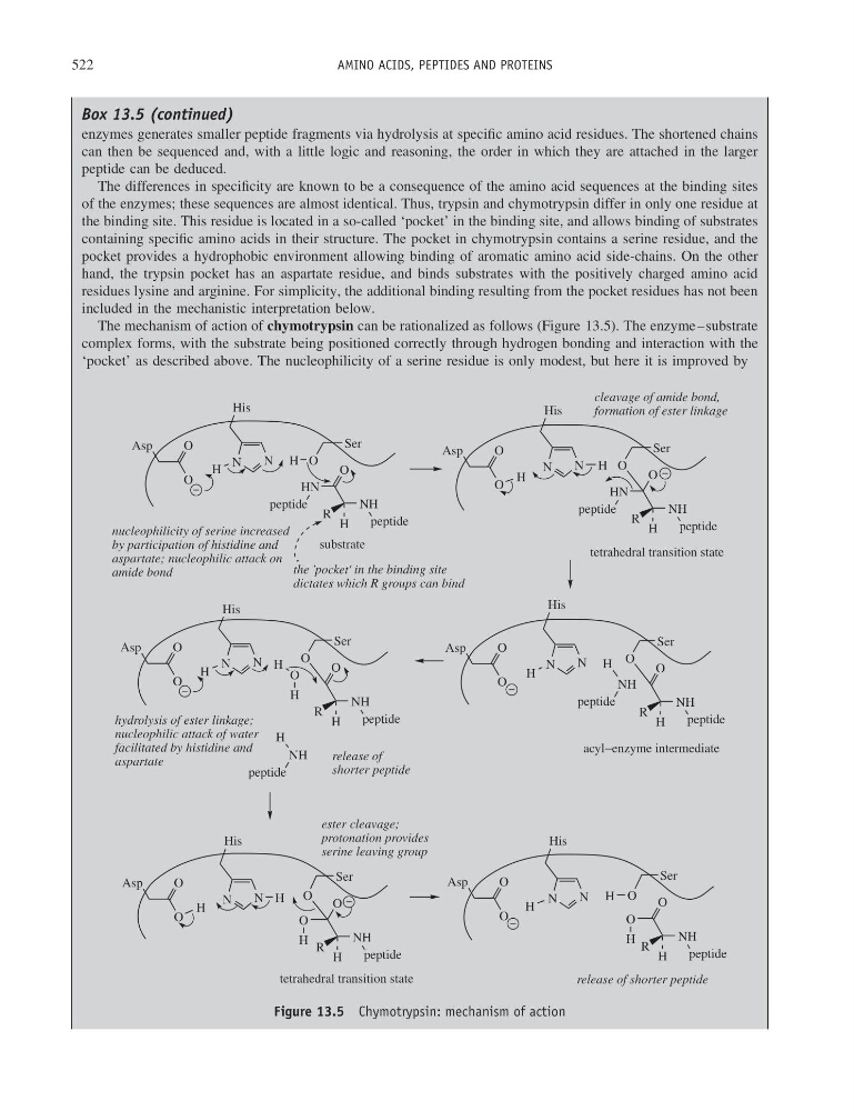
The mechanism of action of chymotrypsin can be rationalized as follows (Figure 13.5). The enzyme–substrate
complex forms, with the substrate being positioned correctly through hydrogen bonding and interaction with the
‘pocket’ as described above. The nucleophilicity of a serine residue is only modest, but here it is improved by
N
N
His
H
O
O
Asp
Ser
O
H
HN
O
peptide
NH
H
R
peptide
N
N
His
H
O
O
Asp
Ser
O
H
HN
O
peptide
NH
H
R
peptide
N
N
His
H
O
O
Asp
Ser
O
H
NH
O
peptide
NH
H
R
peptide
H
NH
peptide
substrate
tetrahedral transition state
acyl−enzyme intermediate
N
N
His
H
O
O
Asp
Ser
O
O
NH
H
R
peptide
H
O
H
N
N
His
H
O
O
Asp
Ser
O
O
NH
H
R
peptide
H
O
H
nucleophilicity of serine increased
by participation of histidine and
aspartate; nucleophilic attack on
amide bond
cleavage of amide bond,
formation of ester linkage
hydrolysis of ester linkage;
nucleophilic attack of water
facilitated by histidine and
aspartate
release of
shorter peptide
ester cleavage;
protonation provides
serine leaving group
release of shorter peptide
N
N
His
H
O
O
Asp
Ser
O
O
NH
H
R
peptide
H
O
H
the 'pocket' in the binding site
dictates which R groups can bind
tetrahedral transition state
Figure 13.5 Chymotrypsin: mechanism of action
AMINO ACIDS, PEPTIDES AND PROTEINS
Box 13.5 (continued)
enzymes generates smaller peptide fragments via hydrolysis at specific amino acid residues. The shortened chains
can then be sequenced and, with a little logic and reasoning, the order in which they are attached in the larger
peptide can be deduced.
The differences in specificity are known to be a consequence of the amino acid sequences at the binding sites
of the enzymes; these sequences are almost identical. Thus, trypsin and chymotrypsin differ in only one residue at
the binding site. This residue is located in a so-called ‘pocket’ in the binding site, and allows binding of substrates
containing specific amino acids in their structure. The pocket in chymotrypsin contains a serine residue, and the
pocket provides a hydrophobic environment allowing binding of aromatic amino acid side-chains. On the other
hand, the trypsin pocket has an aspartate residue, and binds substrates with the positively charged amino acid
residues lysine and arginine. For simplicity, the additional binding resulting from the pocket residues has not been
included in the mechanistic interpretation below.
The mechanism of action of chymotrypsin can be rationalized as follows (Figure 13.5). The enzyme–substrate
complex forms, with the substrate being positioned correctly through hydrogen bonding and interaction with the
‘pocket’ as described above. The nucleophilicity of a serine residue is only modest, but here it is improved by
N
N
His
H
O
O
Asp
Ser
O
H
HN
O
peptide
NH
H
R
peptide
N
N
His
H
O
O
Asp
Ser
O
H
HN
O
peptide
NH
H
R
peptide
N
N
His
H
O
O
Asp
Ser
O
H
NH
O
peptide
NH
H
R
peptide
H
NH
peptide
substrate
tetrahedral transition state
acyl−enzyme intermediate
N
N
His
H
O
O
Asp
Ser
O
O
NH
H
R
peptide
H
O
H
N
N
His
H
O
O
Asp
Ser
O
O
NH
H
R
peptide
H
O
H
nucleophilicity of serine increased
by participation of histidine and
aspartate; nucleophilic attack on
amide bond
cleavage of amide bond,
formation of ester linkage
hydrolysis of ester linkage;
nucleophilic attack of water
facilitated by histidine and
aspartate
release of
shorter peptide
ester cleavage;
protonation provides
serine leaving group
release of shorter peptide
N
N
His
H
O
O
Asp
Ser
O
O
NH
H
R
peptide
H
O
H
the 'pocket' in the binding site
dictates which R groups can bind
tetrahedral transition state
Figure 13.5 Chymotrypsin: mechanism of action