

270
9.3.5 SEM (Scanning Electron Microscopy) Analysis
Scanning electron microscopy is performed in order to obtain important information regarding the biogenetic origin of the raw material and its morphological characteristics. The results of the scanning electron microscopy of the clayey diatomite
are shown in Fig. 9.5. The fragments from the different skeletal forms of microfossils are clearly visible as well as the large number of pores present in the raw material. The sizes of the pores found in the clayey diatomite are in the range 310–650 nm.
9.3.6 TEM Investigations
Transmission electron microscopy results (similarly to SEM) show the presence of
the various microrelics and their morphology. TEM images show globular structures from opal nature with biogenetic origin and relics with different elongated
shapes and other shapes which are most likely due to the clay minerals present in the
sample (Fig. 9.6).
Temp Cel
800.0
600.0
400.0
200.0
DTA uV
40.00
20.00
0.00
-20.00
-40.00
-60.00
-80.00
TG %
100.0
98.0
96.0
94.0
92.0
90.0
88.0
86.0
DTG %/Cel
0.090
0.080
0.070
0.060
0.050
0.040
0.030
0.020
0.010
0.000
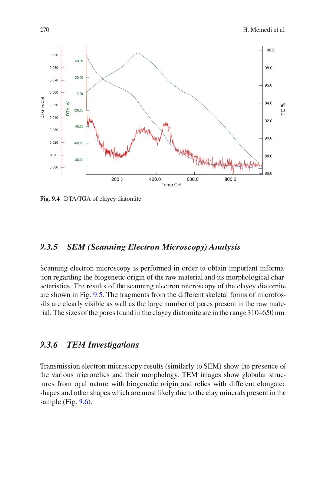
Fig. 9.4 DTA/TGA of clayey diatomite
H. Memedi et al.
9.3.5 SEM (Scanning Electron Microscopy) Analysis
Scanning electron microscopy is performed in order to obtain important information regarding the biogenetic origin of the raw material and its morphological characteristics. The results of the scanning electron microscopy of the clayey diatomite
are shown in Fig. 9.5. The fragments from the different skeletal forms of microfossils are clearly visible as well as the large number of pores present in the raw material. The sizes of the pores found in the clayey diatomite are in the range 310–650 nm.
9.3.6 TEM Investigations
Transmission electron microscopy results (similarly to SEM) show the presence of
the various microrelics and their morphology. TEM images show globular structures from opal nature with biogenetic origin and relics with different elongated
shapes and other shapes which are most likely due to the clay minerals present in the
sample (Fig. 9.6).
Temp Cel
800.0
600.0
400.0
200.0
DTA uV
40.00
20.00
0.00
-20.00
-40.00
-60.00
-80.00
TG %
100.0
98.0
96.0
94.0
92.0
90.0
88.0
86.0
DTG %/Cel
0.090
0.080
0.070
0.060
0.050
0.040
0.030
0.020
0.010
0.000
Fig. 9.4 DTA/TGA of clayey diatomite
H. Memedi et al.