

inflammation and a very strong source of infection. As long as the biofilm exists at
the wound bed, neutrophils and other immune cells accumulate, but the biofilm itself
cannot be destroyed. Therefore, inflammation is sustained due to the endless production of proteases and reactive oxygen species, and healing is impaired. Therefore,
since the latter half of the 2000s, it has been proposed that wound treatment be
selected depending on the presence or absence of biofilm as biofilm-based wound
therapy. However, it is stated that tissue biopsy, observation with an electron
microscope, and observation after special staining are necessary to determine the
presence of a biofilm, and it can be said that implementation in general clinical
practice is difficult. Moreover, in the case of tissue biopsy, since only a small part of
the wound is collected, it is impossible to know how two-dimensionally the biofilm
is distributed. In that sense, conventional biofilm detection technology has limited
clinical applications.
Therefore, we came up with the idea that the components of the biofilm distributed on the wound surface should be extracted and stained. “Wound blotting,” a
technology that can analyze a small amount of protein on the wound surface by
staining the membrane by simply pressing a filter paper called a blotting membrane
on the wound surface for 10 s, can be routinely performed clinically (Minematsu
et al., 2013). We thought that it would be possible to apply this technology to biofilm
detection. This makes it possible to determine not only the presence or absence of the
biofilm but also which part of the wound surface has the biofilm.
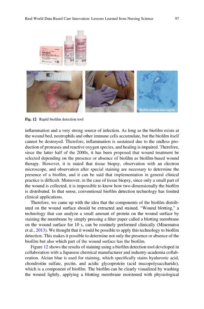
Figure 12 shows the results of staining using a biofilm detection tool developed in
collaboration with a Japanese chemical manufacturer and industry-academia collaboration. Alcian blue is used for staining, which specifically stains hyaluronic acid,
chondroitin sulfate, pectin, and acidic glycoprotein (acid mucopolysaccharide),
which is a component of biofilm. The biofilm can be clearly visualized by washing
the wound lightly, applying a blotting membrane moistened with physiological
Fig. 12 Rapid biofilm detection tool
Real-World Data-Based Care Innovation: Lessons Learned from Nursing Science
97
the wound bed, neutrophils and other immune cells accumulate, but the biofilm itself
cannot be destroyed. Therefore, inflammation is sustained due to the endless production of proteases and reactive oxygen species, and healing is impaired. Therefore,
since the latter half of the 2000s, it has been proposed that wound treatment be
selected depending on the presence or absence of biofilm as biofilm-based wound
therapy. However, it is stated that tissue biopsy, observation with an electron
microscope, and observation after special staining are necessary to determine the
presence of a biofilm, and it can be said that implementation in general clinical
practice is difficult. Moreover, in the case of tissue biopsy, since only a small part of
the wound is collected, it is impossible to know how two-dimensionally the biofilm
is distributed. In that sense, conventional biofilm detection technology has limited
clinical applications.
Therefore, we came up with the idea that the components of the biofilm distributed on the wound surface should be extracted and stained. “Wound blotting,” a
technology that can analyze a small amount of protein on the wound surface by
staining the membrane by simply pressing a filter paper called a blotting membrane
on the wound surface for 10 s, can be routinely performed clinically (Minematsu
et al., 2013). We thought that it would be possible to apply this technology to biofilm
detection. This makes it possible to determine not only the presence or absence of the
biofilm but also which part of the wound surface has the biofilm.
Figure 12 shows the results of staining using a biofilm detection tool developed in
collaboration with a Japanese chemical manufacturer and industry-academia collaboration. Alcian blue is used for staining, which specifically stains hyaluronic acid,
chondroitin sulfate, pectin, and acidic glycoprotein (acid mucopolysaccharide),
which is a component of biofilm. The biofilm can be clearly visualized by washing
the wound lightly, applying a blotting membrane moistened with physiological
Fig. 12 Rapid biofilm detection tool
Real-World Data-Based Care Innovation: Lessons Learned from Nursing Science
97