

with the exception of Feramyces and Ghazallomyces, form monoflagellate zoospores.
Culture-independent diversity surveys based
on the DNA of the internal transcribed spacer
region (ITS) did suggest that additional clades
were present, and the first DNA metabarcoding
study of anaerobic fungi (Liggenstoffer et al.
2010) identified the existence of several novel
clades, some of which have subsequently been
isolated into pure cultures (Fig. 17.3). Due to
the high level of intragenomic variability in the
ITS region, the adjacent large ribosomal subunit (LSU; D1/D2 variable regions; Fig. 17.3) is
now currently favored for accurate phylogenetic reconstructions (Dagar et al. 2011).
Although the niche of anaerobic fungi is the
digestive tract of large mammalian herbivores,
most laboratory isolates have been obtained
from herbivore feces. Moreover, fungi in feces
and post-rumen digesta remain viable when
dried in air at ambient temperature (Davies
et al. 1993) and even in sun-baked feces
(Milne et al. 1989). Although populations tend
to decline slowly after drying, the size of the
anaerobic fungal community in fresh feces,
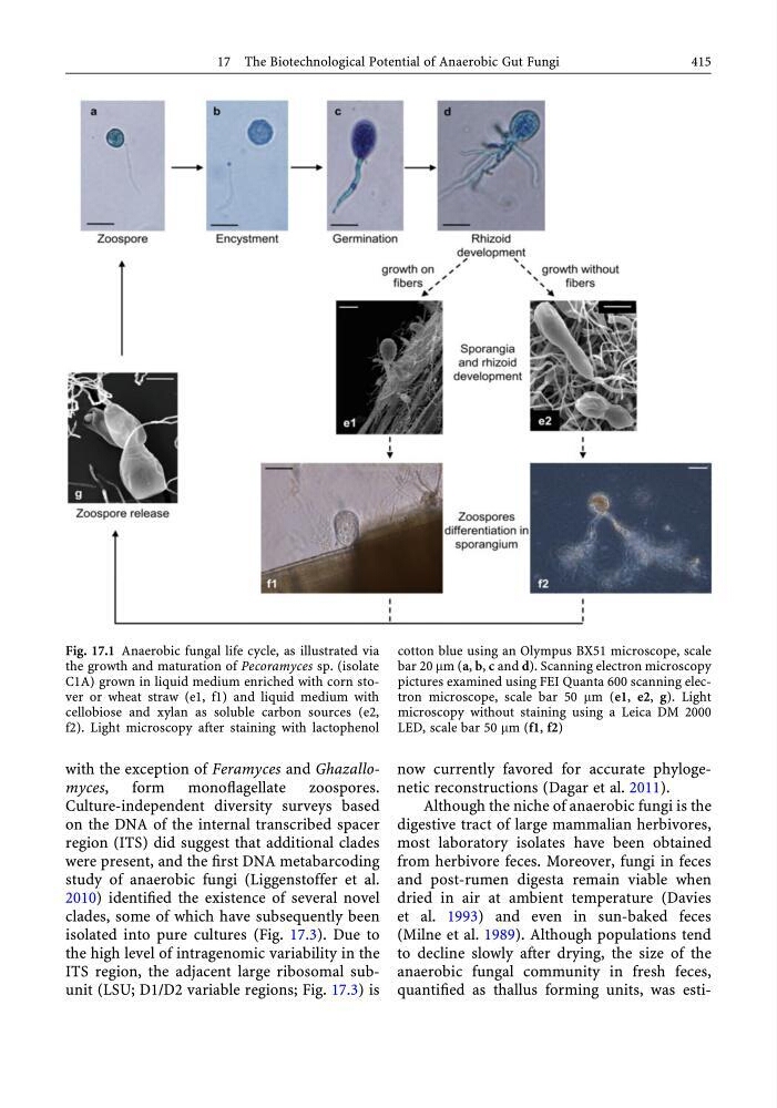
quantified as thallus forming units, was estiFig. 17.1 Anaerobic fungal life cycle, as illustrated via
the growth and maturation of Pecoramyces sp. (isolate
C1A) grown in liquid medium enriched with corn stover or wheat straw (e1, f1) and liquid medium with
cellobiose and xylan as soluble carbon sources (e2,
f2). Light microscopy after staining with lactophenol
cotton blue using an Olympus BX51 microscope, scale
bar 20 mm (a, b, c and d). Scanning electron microscopy
pictures examined using FEI Quanta 600 scanning electron microscope, scale bar 50 mm (e1, e2, g). Light
microscopy without staining using a Leica DM 2000
LED, scale bar 50 mm (f1, f2)
17 The Biotechnological Potential of Anaerobic Gut Fungi
415
Culture-independent diversity surveys based
on the DNA of the internal transcribed spacer
region (ITS) did suggest that additional clades
were present, and the first DNA metabarcoding
study of anaerobic fungi (Liggenstoffer et al.
2010) identified the existence of several novel
clades, some of which have subsequently been
isolated into pure cultures (Fig. 17.3). Due to
the high level of intragenomic variability in the
ITS region, the adjacent large ribosomal subunit (LSU; D1/D2 variable regions; Fig. 17.3) is
now currently favored for accurate phylogenetic reconstructions (Dagar et al. 2011).
Although the niche of anaerobic fungi is the
digestive tract of large mammalian herbivores,
most laboratory isolates have been obtained
from herbivore feces. Moreover, fungi in feces
and post-rumen digesta remain viable when
dried in air at ambient temperature (Davies
et al. 1993) and even in sun-baked feces
(Milne et al. 1989). Although populations tend
to decline slowly after drying, the size of the
anaerobic fungal community in fresh feces,
quantified as thallus forming units, was estiFig. 17.1 Anaerobic fungal life cycle, as illustrated via
the growth and maturation of Pecoramyces sp. (isolate
C1A) grown in liquid medium enriched with corn stover or wheat straw (e1, f1) and liquid medium with
cellobiose and xylan as soluble carbon sources (e2,
f2). Light microscopy after staining with lactophenol
cotton blue using an Olympus BX51 microscope, scale
bar 20 mm (a, b, c and d). Scanning electron microscopy
pictures examined using FEI Quanta 600 scanning electron microscope, scale bar 50 mm (e1, e2, g). Light
microscopy without staining using a Leica DM 2000
LED, scale bar 50 mm (f1, f2)
17 The Biotechnological Potential of Anaerobic Gut Fungi
415