

58
M. Stein
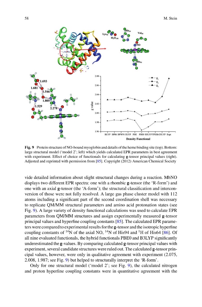
Fig. 9 Protein structure of NO-bound myoglobin and details of the heme binding site (top). Bottom:
large structural model (‘model 2’; left) which yields calculated EPR parameters in best agreement
with experiment. Effect of choice of functionals for calculating g-tensor principal values (right).
Adjusted and reprinted with permission from [85]. Copyright (2012) American Chemical Society
vide detailed information about slight structural changes during a reaction. MbNO
displays two different EPR spectra: one with a rhombic g-tensor (the ‘R-form’) and
one with an axial g-tensor (the ‘A-form’); the structural classification and interconversion of those were not fully resolved. A large gas phase cluster model with 112
atoms including a significant part of the second coordination shell was necessary
to replicate QM/MM structural parameters and amino acid protonation states (see
Fig. 9). A large variety of density functional calculations was used to calculate EPR
parameters from QM/MM structures and assign experimentally measured g-tensor
principal values and hyperfine coupling constants [85]. The calculated EPR parameters were compared to experimental results for the g-tensor and the isotropic hyperfine
coupling constants of
14 N of the axial NO,
14 N of His94 and
1 H of His64 [86]. Of
all nine evaluated functionals, the hybrid functionals PBE0 and B3LYP significantly
underestimated the g-values. By comparing calculated g-tensor principal values with
experiment, several candidate structures were ruled out. The calculated g-tensor principal values, however, were only in qualitative agreement with experiment (2.075,
2.008, 1.987; see Fig. 9) but helped to structurally interpret the ‘R-form’.
Only for one structural model (‘model 2’; see Fig. 9), the calculated nitrogen
and proton hyperfine coupling constants were in quantitative agreement with the
M. Stein
Fig. 9 Protein structure of NO-bound myoglobin and details of the heme binding site (top). Bottom:
large structural model (‘model 2’; left) which yields calculated EPR parameters in best agreement
with experiment. Effect of choice of functionals for calculating g-tensor principal values (right).
Adjusted and reprinted with permission from [85]. Copyright (2012) American Chemical Society
vide detailed information about slight structural changes during a reaction. MbNO
displays two different EPR spectra: one with a rhombic g-tensor (the ‘R-form’) and
one with an axial g-tensor (the ‘A-form’); the structural classification and interconversion of those were not fully resolved. A large gas phase cluster model with 112
atoms including a significant part of the second coordination shell was necessary
to replicate QM/MM structural parameters and amino acid protonation states (see
Fig. 9). A large variety of density functional calculations was used to calculate EPR
parameters from QM/MM structures and assign experimentally measured g-tensor
principal values and hyperfine coupling constants [85]. The calculated EPR parameters were compared to experimental results for the g-tensor and the isotropic hyperfine
coupling constants of
14 N of the axial NO,
14 N of His94 and
1 H of His64 [86]. Of
all nine evaluated functionals, the hybrid functionals PBE0 and B3LYP significantly
underestimated the g-values. By comparing calculated g-tensor principal values with
experiment, several candidate structures were ruled out. The calculated g-tensor principal values, however, were only in qualitative agreement with experiment (2.075,
2.008, 1.987; see Fig. 9) but helped to structurally interpret the ‘R-form’.
Only for one structural model (‘model 2’; see Fig. 9), the calculated nitrogen
and proton hyperfine coupling constants were in quantitative agreement with the