

Anisotropic Magnetic Spin Interactions of Transition Metal …
57
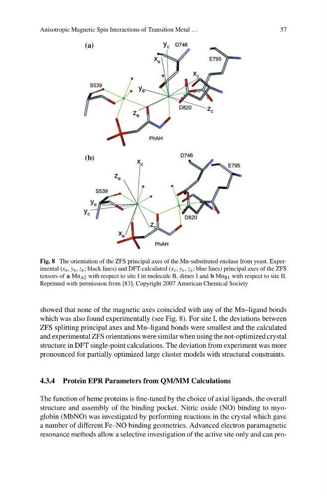
Fig. 8 The orientation of the ZFS principal axes of the Mn-substituted enolase from yeast. Experimental (x e , y e , z e ; black lines) and DFT-calculated (x c , y c , z c ; blue lines) principal axes of the ZFS
tensors of a Mn A2 with respect to site I in molecule B, dimer I and b Mn B1 with respect to site II.
Reprinted with permission from [83]. Copyright 2007 American Chemical Society
showed that none of the magnetic axes coincided with any of the Mn–ligand bonds
which was also found experimentally (see Fig. 8). For site I, the deviations between
ZFS splitting principal axes and Mn–ligand bonds were smallest and the calculated
and experimental ZFS orientations were similar when using the not-optimized crystal
structure in DFT single-point calculations. The deviation from experiment was more
pronounced for partially optimized large cluster models with structural constraints.
4.3.4 Protein EPR Parameters from QM/MM Calculations
The function of heme proteins is fine-tuned by the choice of axial ligands, the overall
structure and assembly of the binding pocket. Nitric oxide (NO) binding to myoglobin (MbNO) was investigated by performing reactions in the crystal which gave
a number of different Fe–NO binding geometries. Advanced electron paramagnetic
resonance methods allow a selective investigation of the active site only and can pro-
57
Fig. 8 The orientation of the ZFS principal axes of the Mn-substituted enolase from yeast. Experimental (x e , y e , z e ; black lines) and DFT-calculated (x c , y c , z c ; blue lines) principal axes of the ZFS
tensors of a Mn A2 with respect to site I in molecule B, dimer I and b Mn B1 with respect to site II.
Reprinted with permission from [83]. Copyright 2007 American Chemical Society
showed that none of the magnetic axes coincided with any of the Mn–ligand bonds
which was also found experimentally (see Fig. 8). For site I, the deviations between
ZFS splitting principal axes and Mn–ligand bonds were smallest and the calculated
and experimental ZFS orientations were similar when using the not-optimized crystal
structure in DFT single-point calculations. The deviation from experiment was more
pronounced for partially optimized large cluster models with structural constraints.
4.3.4 Protein EPR Parameters from QM/MM Calculations
The function of heme proteins is fine-tuned by the choice of axial ligands, the overall
structure and assembly of the binding pocket. Nitric oxide (NO) binding to myoglobin (MbNO) was investigated by performing reactions in the crystal which gave
a number of different Fe–NO binding geometries. Advanced electron paramagnetic
resonance methods allow a selective investigation of the active site only and can pro-