

54
M. Stein
crystal EPR investigations and a definite assignment of the respective paramagnetic
site in the unit cell states from angle-dependent EPR spectra was feasible [78, 80].
In the high field approximation, the orientation-dependent hyperfine splitting is
observed in the principal axes system x, y, z of the hyperfine tensor A
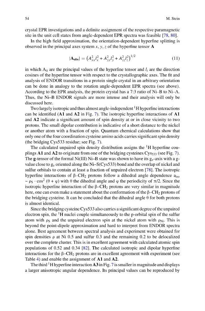
|A obs |
A
2
xx l
2
x + A
2
yy l
2
y + A
2
zz l
2
z
1/2
(11)
in which A ii are the principal values of the hyperfine tensor and l i are the direction
cosines of the hyperfine tensor with respect to the crystallographic axes. The fit and
analysis of ENDOR transitions in a protein single crystal in an arbitrary orientation
can be done in analogy to the rotation angle-dependent EPR spectra (see above).
According to the EPR analysis, the protein crystal has a 7:3 ratio of Ni–B to Ni–A.
Thus, the Ni–B ENDOR signals are more intense and their analysis will only be
discussed here.
Two largely isotropic and thus almost angle-independent
1 H hyperfine interactions
can be identified (A1 and A2 in Fig. 7). The isotropic hyperfine interactions of A1
and A2 indicate a significant amount of spin density at or in close vicinity to two
protons. The small dipolar contribution is indicative of a short distance to the nickel
or another atom with a fraction of spin. Quantum chemical calculations show that
only one of the four coordination cysteine amino acids carries significant spin density
(the bridging Cys533 residue; see Fig. 7).
The calculated unpaired spin density distribution assigns the
1 H hyperfine couplings A1 and A2 to originate from one of the bridging cysteines Cys 533 (see Fig. 7).
The g-tensor of the formal Ni(III) Ni–B state was shown to have its g z -axis with a gvalue close to g e oriented along the Ni–S(Cys533) bond and the overlap of nickel and
sulfur orbitals to contain at least a fraction of unpaired electron [78]. The isotropic
hyperfine interactions of β–CH 2 protons follow a dihedral angle dependence a iso
~ ρ S · cos
2 (θ + ϕ) with θ the dihedral angle and ϕ the periodicity of π/2. Since the
isotropic hyperfine interaction of the β–CH 2 protons are very similar in magnitude
here, one can even make a statement about the conformation of the β–CH 2 protons of
the bridging cysteine. It can be concluded that the dihedral angle θ for both protons
is almost identical.
Since the bridging cysteine Cys533 also carries a significant degree of the unpaired
electron spin, the
1 H nuclei couple simultaneously to the p-orbital spin of the sulfur
atom with ρ S and the unpaired electron spin at the nickel atom with ρ Ni . This is
beyond the point-dipole approximation and hard to interpret from ENDOR spectra
alone. Best agreement between spectral analysis and experiment were obtained for
spin densities ρ at Ni 0.5 and sulfur 0.3 and the remaining 0.2 to be delocalized
over the complete cluster. This is in excellent agreement with calculated atomic spin
populations of 0.52 and 0.34 [82]. The calculated isotropic and dipolar hyperfine
interactions for the β–CH 2 protons are in excellent agreement with experiment (see
Table 4) and enable the assignment of A1 and A2.
The third
1 H hyperfine interaction A3 in Fig. 7 is smaller in magnitude and displays
a larger anisotropic angular dependence. Its principal values can be reproduced by
M. Stein
crystal EPR investigations and a definite assignment of the respective paramagnetic
site in the unit cell states from angle-dependent EPR spectra was feasible [78, 80].
In the high field approximation, the orientation-dependent hyperfine splitting is
observed in the principal axes system x, y, z of the hyperfine tensor A
|A obs |
A
2
xx l
2
x + A
2
yy l
2
y + A
2
zz l
2
z
1/2
(11)
in which A ii are the principal values of the hyperfine tensor and l i are the direction
cosines of the hyperfine tensor with respect to the crystallographic axes. The fit and
analysis of ENDOR transitions in a protein single crystal in an arbitrary orientation
can be done in analogy to the rotation angle-dependent EPR spectra (see above).
According to the EPR analysis, the protein crystal has a 7:3 ratio of Ni–B to Ni–A.
Thus, the Ni–B ENDOR signals are more intense and their analysis will only be
discussed here.
Two largely isotropic and thus almost angle-independent
1 H hyperfine interactions
can be identified (A1 and A2 in Fig. 7). The isotropic hyperfine interactions of A1
and A2 indicate a significant amount of spin density at or in close vicinity to two
protons. The small dipolar contribution is indicative of a short distance to the nickel
or another atom with a fraction of spin. Quantum chemical calculations show that
only one of the four coordination cysteine amino acids carries significant spin density
(the bridging Cys533 residue; see Fig. 7).
The calculated unpaired spin density distribution assigns the
1 H hyperfine couplings A1 and A2 to originate from one of the bridging cysteines Cys 533 (see Fig. 7).
The g-tensor of the formal Ni(III) Ni–B state was shown to have its g z -axis with a gvalue close to g e oriented along the Ni–S(Cys533) bond and the overlap of nickel and
sulfur orbitals to contain at least a fraction of unpaired electron [78]. The isotropic
hyperfine interactions of β–CH 2 protons follow a dihedral angle dependence a iso
~ ρ S · cos
2 (θ + ϕ) with θ the dihedral angle and ϕ the periodicity of π/2. Since the
isotropic hyperfine interaction of the β–CH 2 protons are very similar in magnitude
here, one can even make a statement about the conformation of the β–CH 2 protons of
the bridging cysteine. It can be concluded that the dihedral angle θ for both protons
is almost identical.
Since the bridging cysteine Cys533 also carries a significant degree of the unpaired
electron spin, the
1 H nuclei couple simultaneously to the p-orbital spin of the sulfur
atom with ρ S and the unpaired electron spin at the nickel atom with ρ Ni . This is
beyond the point-dipole approximation and hard to interpret from ENDOR spectra
alone. Best agreement between spectral analysis and experiment were obtained for
spin densities ρ at Ni 0.5 and sulfur 0.3 and the remaining 0.2 to be delocalized
over the complete cluster. This is in excellent agreement with calculated atomic spin
populations of 0.52 and 0.34 [82]. The calculated isotropic and dipolar hyperfine
interactions for the β–CH 2 protons are in excellent agreement with experiment (see
Table 4) and enable the assignment of A1 and A2.
The third
1 H hyperfine interaction A3 in Fig. 7 is smaller in magnitude and displays
a larger anisotropic angular dependence. Its principal values can be reproduced by