

Challenges in Modelling Metalloenzymes
511
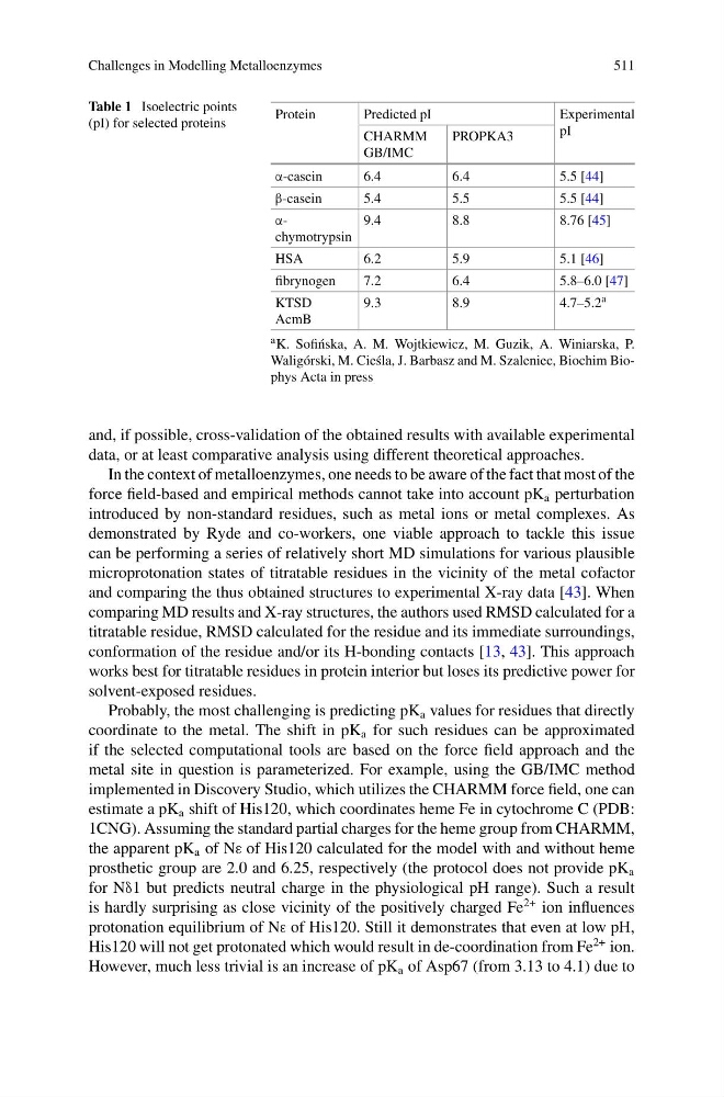
Table 1 Isoelectric points
(pI) for selected proteins
Protein
Predicted pI
Experimental
pI
CHARMM
GB/IMC
PROPKA3
α-casein
6.4
6.4
5.5 [44]
β-casein
5.4
5.5
5.5 [44]
αchymotrypsin
9.4
8.8
8.76 [45]
HSA
6.2
5.9
5.1 [46]
fibrynogen
7.2
6.4
5.8–6.0 [47]
KTSD
AcmB
9.3
8.9
4.7–5.2 a
a K. Sofi´ nska, A. M. Wojtkiewicz, M. Guzik, A. Winiarska, P.
Waligórski, M. Cie´ sla, J. Barbasz and M. Szaleniec, Biochim Biophys Acta in press
and, if possible, cross-validation of the obtained results with available experimental
data, or at least comparative analysis using different theoretical approaches.
In the context of metalloenzymes, one needs to be aware of the fact that most of the
force field-based and empirical methods cannot take into account pK a perturbation
introduced by non-standard residues, such as metal ions or metal complexes. As
demonstrated by Ryde and co-workers, one viable approach to tackle this issue
can be performing a series of relatively short MD simulations for various plausible
microprotonation states of titratable residues in the vicinity of the metal cofactor
and comparing the thus obtained structures to experimental X-ray data [43]. When
comparing MD results and X-ray structures, the authors used RMSD calculated for a
titratable residue, RMSD calculated for the residue and its immediate surroundings,
conformation of the residue and/or its H-bonding contacts [13, 43]. This approach
works best for titratable residues in protein interior but loses its predictive power for
solvent-exposed residues.
Probably, the most challenging is predicting pK a values for residues that directly
coordinate to the metal. The shift in pK a for such residues can be approximated
if the selected computational tools are based on the force field approach and the
metal site in question is parameterized. For example, using the GB/IMC method
implemented in Discovery Studio, which utilizes the CHARMM force field, one can
estimate a pK a shift of His120, which coordinates heme Fe in cytochrome C (PDB:
1CNG). Assuming the standard partial charges for the heme group from CHARMM,
the apparent pK a of Nε of His120 calculated for the model with and without heme
prosthetic group are 2.0 and 6.25, respectively (the protocol does not provide pK a
for Nδ1 but predicts neutral charge in the physiological pH range). Such a result
is hardly surprising as close vicinity of the positively charged Fe
2+ ion influences
protonation equilibrium of Nε of His120. Still it demonstrates that even at low pH,
His120 will not get protonated which would result in de-coordination from Fe
2+ ion.
However, much less trivial is an increase of pK a of Asp67 (from 3.13 to 4.1) due to
511
Table 1 Isoelectric points
(pI) for selected proteins
Protein
Predicted pI
Experimental
pI
CHARMM
GB/IMC
PROPKA3
α-casein
6.4
6.4
5.5 [44]
β-casein
5.4
5.5
5.5 [44]
αchymotrypsin
9.4
8.8
8.76 [45]
HSA
6.2
5.9
5.1 [46]
fibrynogen
7.2
6.4
5.8–6.0 [47]
KTSD
AcmB
9.3
8.9
4.7–5.2 a
a K. Sofi´ nska, A. M. Wojtkiewicz, M. Guzik, A. Winiarska, P.
Waligórski, M. Cie´ sla, J. Barbasz and M. Szaleniec, Biochim Biophys Acta in press
and, if possible, cross-validation of the obtained results with available experimental
data, or at least comparative analysis using different theoretical approaches.
In the context of metalloenzymes, one needs to be aware of the fact that most of the
force field-based and empirical methods cannot take into account pK a perturbation
introduced by non-standard residues, such as metal ions or metal complexes. As
demonstrated by Ryde and co-workers, one viable approach to tackle this issue
can be performing a series of relatively short MD simulations for various plausible
microprotonation states of titratable residues in the vicinity of the metal cofactor
and comparing the thus obtained structures to experimental X-ray data [43]. When
comparing MD results and X-ray structures, the authors used RMSD calculated for a
titratable residue, RMSD calculated for the residue and its immediate surroundings,
conformation of the residue and/or its H-bonding contacts [13, 43]. This approach
works best for titratable residues in protein interior but loses its predictive power for
solvent-exposed residues.
Probably, the most challenging is predicting pK a values for residues that directly
coordinate to the metal. The shift in pK a for such residues can be approximated
if the selected computational tools are based on the force field approach and the
metal site in question is parameterized. For example, using the GB/IMC method
implemented in Discovery Studio, which utilizes the CHARMM force field, one can
estimate a pK a shift of His120, which coordinates heme Fe in cytochrome C (PDB:
1CNG). Assuming the standard partial charges for the heme group from CHARMM,
the apparent pK a of Nε of His120 calculated for the model with and without heme
prosthetic group are 2.0 and 6.25, respectively (the protocol does not provide pK a
for Nδ1 but predicts neutral charge in the physiological pH range). Such a result
is hardly surprising as close vicinity of the positively charged Fe
2+ ion influences
protonation equilibrium of Nε of His120. Still it demonstrates that even at low pH,
His120 will not get protonated which would result in de-coordination from Fe
2+ ion.
However, much less trivial is an increase of pK a of Asp67 (from 3.13 to 4.1) due to