

Applications of Computational Chemistry to Selected Problems …
475
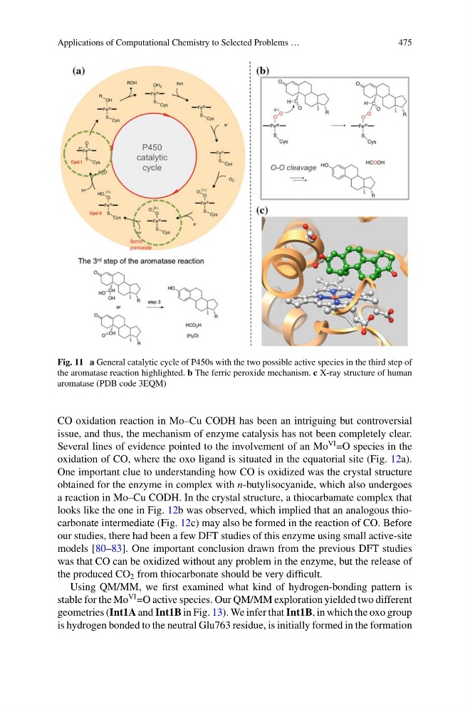
Fig. 11 a General catalytic cycle of P450s with the two possible active species in the third step of
the aromatase reaction highlighted. b The ferric peroxide mechanism. c X-ray structure of human
aromatase (PDB code 3EQM)
CO oxidation reaction in Mo–Cu CODH has been an intriguing but controversial
issue, and thus, the mechanism of enzyme catalysis has not been completely clear.
Several lines of evidence pointed to the involvement of an Mo
VI =O species in the
oxidation of CO, where the oxo ligand is situated in the equatorial site (Fig. 12a).
One important clue to understanding how CO is oxidized was the crystal structure
obtained for the enzyme in complex with n-butylisocyanide, which also undergoes
a reaction in Mo–Cu CODH. In the crystal structure, a thiocarbamate complex that
looks like the one in Fig. 12b was observed, which implied that an analogous thiocarbonate intermediate (Fig. 12c) may also be formed in the reaction of CO. Before
our studies, there had been a few DFT studies of this enzyme using small active-site
models [80–83]. One important conclusion drawn from the previous DFT studies
was that CO can be oxidized without any problem in the enzyme, but the release of
the produced CO 2 from thiocarbonate should be very difficult.
Using QM/MM, we first examined what kind of hydrogen-bonding pattern is
stable for the Mo
VI =O active species. Our QM/MM exploration yielded two different
geometries (Int1A and Int1B in Fig. 13). We infer that Int1B, in which the oxo group
is hydrogen bonded to the neutral Glu763 residue, is initially formed in the formation
475
Fig. 11 a General catalytic cycle of P450s with the two possible active species in the third step of
the aromatase reaction highlighted. b The ferric peroxide mechanism. c X-ray structure of human
aromatase (PDB code 3EQM)
CO oxidation reaction in Mo–Cu CODH has been an intriguing but controversial
issue, and thus, the mechanism of enzyme catalysis has not been completely clear.
Several lines of evidence pointed to the involvement of an Mo
VI =O species in the
oxidation of CO, where the oxo ligand is situated in the equatorial site (Fig. 12a).
One important clue to understanding how CO is oxidized was the crystal structure
obtained for the enzyme in complex with n-butylisocyanide, which also undergoes
a reaction in Mo–Cu CODH. In the crystal structure, a thiocarbamate complex that
looks like the one in Fig. 12b was observed, which implied that an analogous thiocarbonate intermediate (Fig. 12c) may also be formed in the reaction of CO. Before
our studies, there had been a few DFT studies of this enzyme using small active-site
models [80–83]. One important conclusion drawn from the previous DFT studies
was that CO can be oxidized without any problem in the enzyme, but the release of
the produced CO 2 from thiocarbonate should be very difficult.
Using QM/MM, we first examined what kind of hydrogen-bonding pattern is
stable for the Mo
VI =O active species. Our QM/MM exploration yielded two different
geometries (Int1A and Int1B in Fig. 13). We infer that Int1B, in which the oxo group
is hydrogen bonded to the neutral Glu763 residue, is initially formed in the formation