

Assessing Electronically Excited States of Cobalamins …
249
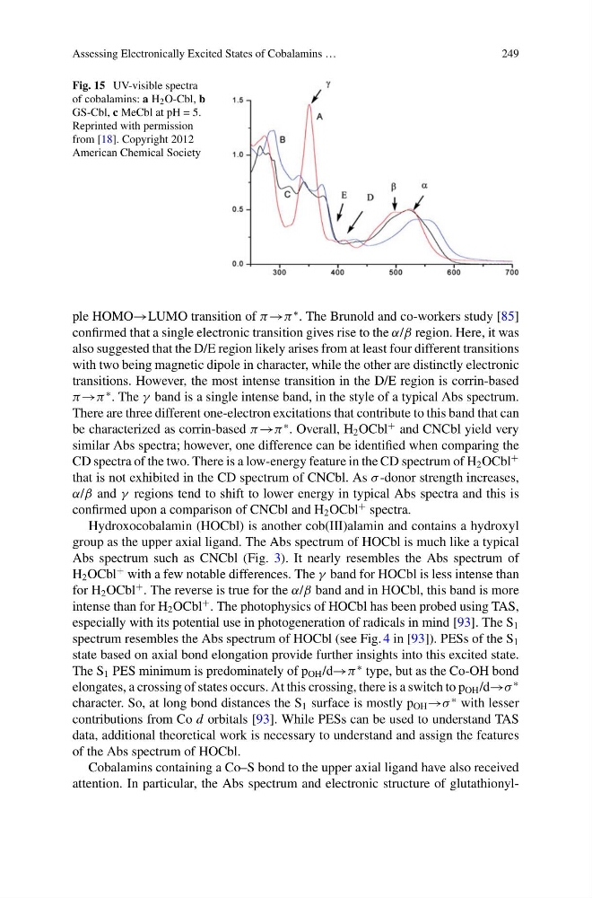
Fig. 15 UV-visible spectra
of cobalamins: a H 2 O-Cbl, b
GS-Cbl, c MeCbl at pH = 5.
Reprinted with permission
from [18]. Copyright 2012
American Chemical Society
ple HOMO→LUMO transition of π →π
∗ . The Brunold and co-workers study [85]
confirmed that a single electronic transition gives rise to the α/β region. Here, it was
also suggested that the D/E region likely arises from at least four different transitions
with two being magnetic dipole in character, while the other are distinctly electronic
transitions. However, the most intense transition in the D/E region is corrin-based
π →π
∗ . The γ band is a single intense band, in the style of a typical Abs spectrum.
There are three different one-electron excitations that contribute to this band that can
be characterized as corrin-based π →π
∗ . Overall, H 2 OCbl
+ and CNCbl yield very
similar Abs spectra; however, one difference can be identified when comparing the
CD spectra of the two. There is a low-energy feature in the CD spectrum of H 2 OCbl
+
that is not exhibited in the CD spectrum of CNCbl. As σ -donor strength increases,
α/β and γ regions tend to shift to lower energy in typical Abs spectra and this is
confirmed upon a comparison of CNCbl and H 2 OCbl
+ spectra.
Hydroxocobalamin (HOCbl) is another cob(III)alamin and contains a hydroxyl
group as the upper axial ligand. The Abs spectrum of HOCbl is much like a typical
Abs spectrum such as CNCbl (Fig. 3). It nearly resembles the Abs spectrum of
H 2 OCbl
+ with a few notable differences. The γ band for HOCbl is less intense than
for H 2 OCbl
+ . The reverse is true for the α/β band and in HOCbl, this band is more
intense than for H 2 OCbl
+ . The photophysics of HOCbl has been probed using TAS,
especially with its potential use in photogeneration of radicals in mind [93]. The S 1
spectrum resembles the Abs spectrum of HOCbl (see Fig. 4 in [93]). PESs of the S 1
state based on axial bond elongation provide further insights into this excited state.
The S 1 PES minimum is predominately of p OH /d→π
∗ type, but as the Co-OH bond
elongates, a crossing of states occurs. At this crossing, there is a switch to p OH /d→σ
∗
character. So, at long bond distances the S 1 surface is mostly p OH →σ
∗ with lesser
contributions from Co d orbitals [93]. While PESs can be used to understand TAS
data, additional theoretical work is necessary to understand and assign the features
of the Abs spectrum of HOCbl.
Cobalamins containing a Co–S bond to the upper axial ligand have also received
attention. In particular, the Abs spectrum and electronic structure of glutathionyl-
249
Fig. 15 UV-visible spectra
of cobalamins: a H 2 O-Cbl, b
GS-Cbl, c MeCbl at pH = 5.
Reprinted with permission
from [18]. Copyright 2012
American Chemical Society
ple HOMO→LUMO transition of π →π
∗ . The Brunold and co-workers study [85]
confirmed that a single electronic transition gives rise to the α/β region. Here, it was
also suggested that the D/E region likely arises from at least four different transitions
with two being magnetic dipole in character, while the other are distinctly electronic
transitions. However, the most intense transition in the D/E region is corrin-based
π →π
∗ . The γ band is a single intense band, in the style of a typical Abs spectrum.
There are three different one-electron excitations that contribute to this band that can
be characterized as corrin-based π →π
∗ . Overall, H 2 OCbl
+ and CNCbl yield very
similar Abs spectra; however, one difference can be identified when comparing the
CD spectra of the two. There is a low-energy feature in the CD spectrum of H 2 OCbl
+
that is not exhibited in the CD spectrum of CNCbl. As σ -donor strength increases,
α/β and γ regions tend to shift to lower energy in typical Abs spectra and this is
confirmed upon a comparison of CNCbl and H 2 OCbl
+ spectra.
Hydroxocobalamin (HOCbl) is another cob(III)alamin and contains a hydroxyl
group as the upper axial ligand. The Abs spectrum of HOCbl is much like a typical
Abs spectrum such as CNCbl (Fig. 3). It nearly resembles the Abs spectrum of
H 2 OCbl
+ with a few notable differences. The γ band for HOCbl is less intense than
for H 2 OCbl
+ . The reverse is true for the α/β band and in HOCbl, this band is more
intense than for H 2 OCbl
+ . The photophysics of HOCbl has been probed using TAS,
especially with its potential use in photogeneration of radicals in mind [93]. The S 1
spectrum resembles the Abs spectrum of HOCbl (see Fig. 4 in [93]). PESs of the S 1
state based on axial bond elongation provide further insights into this excited state.
The S 1 PES minimum is predominately of p OH /d→π
∗ type, but as the Co-OH bond
elongates, a crossing of states occurs. At this crossing, there is a switch to p OH /d→σ
∗
character. So, at long bond distances the S 1 surface is mostly p OH →σ
∗ with lesser
contributions from Co d orbitals [93]. While PESs can be used to understand TAS
data, additional theoretical work is necessary to understand and assign the features
of the Abs spectrum of HOCbl.
Cobalamins containing a Co–S bond to the upper axial ligand have also received
attention. In particular, the Abs spectrum and electronic structure of glutathionyl-