

234
M. J. Toda et al.
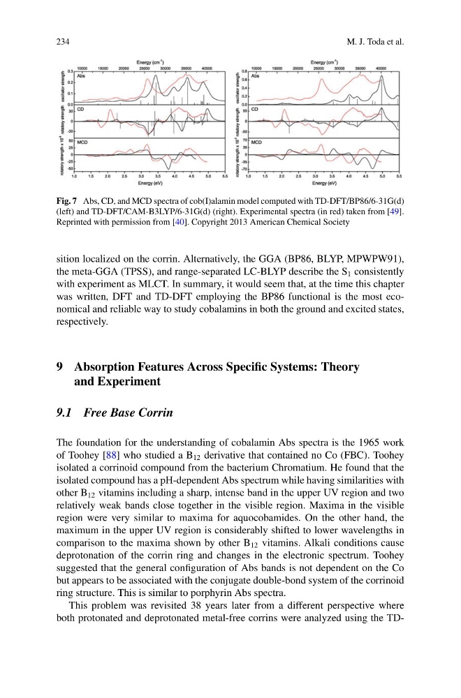
Fig. 7 Abs, CD, and MCD spectra of cob(I)alamin model computed with TD-DFT/BP86/6-31G(d)
(left) and TD-DFT/CAM-B3LYP/6-31G(d) (right). Experimental spectra (in red) taken from [49].
Reprinted with permission from [40]. Copyright 2013 American Chemical Society
sition localized on the corrin. Alternatively, the GGA (BP86, BLYP, MPWPW91),
the meta-GGA (TPSS), and range-separated LC-BLYP describe the S 1 consistently
with experiment as MLCT. In summary, it would seem that, at the time this chapter
was written, DFT and TD-DFT employing the BP86 functional is the most economical and reliable way to study cobalamins in both the ground and excited states,
respectively.
9 Absorption Features Across Specific Systems: Theory
and Experiment
9.1 Free Base Corrin
The foundation for the understanding of cobalamin Abs spectra is the 1965 work
of Toohey [88] who studied a B 12 derivative that contained no Co (FBC). Toohey
isolated a corrinoid compound from the bacterium Chromatium. He found that the
isolated compound has a pH-dependent Abs spectrum while having similarities with
other B 12 vitamins including a sharp, intense band in the upper UV region and two
relatively weak bands close together in the visible region. Maxima in the visible
region were very similar to maxima for aquocobamides. On the other hand, the
maximum in the upper UV region is considerably shifted to lower wavelengths in
comparison to the maxima shown by other B 12 vitamins. Alkali conditions cause
deprotonation of the corrin ring and changes in the electronic spectrum. Toohey
suggested that the general configuration of Abs bands is not dependent on the Co
but appears to be associated with the conjugate double-bond system of the corrinoid
ring structure. This is similar to porphyrin Abs spectra.
This problem was revisited 38 years later from a different perspective where
both protonated and deprotonated metal-free corrins were analyzed using the TD-
M. J. Toda et al.
Fig. 7 Abs, CD, and MCD spectra of cob(I)alamin model computed with TD-DFT/BP86/6-31G(d)
(left) and TD-DFT/CAM-B3LYP/6-31G(d) (right). Experimental spectra (in red) taken from [49].
Reprinted with permission from [40]. Copyright 2013 American Chemical Society
sition localized on the corrin. Alternatively, the GGA (BP86, BLYP, MPWPW91),
the meta-GGA (TPSS), and range-separated LC-BLYP describe the S 1 consistently
with experiment as MLCT. In summary, it would seem that, at the time this chapter
was written, DFT and TD-DFT employing the BP86 functional is the most economical and reliable way to study cobalamins in both the ground and excited states,
respectively.
9 Absorption Features Across Specific Systems: Theory
and Experiment
9.1 Free Base Corrin
The foundation for the understanding of cobalamin Abs spectra is the 1965 work
of Toohey [88] who studied a B 12 derivative that contained no Co (FBC). Toohey
isolated a corrinoid compound from the bacterium Chromatium. He found that the
isolated compound has a pH-dependent Abs spectrum while having similarities with
other B 12 vitamins including a sharp, intense band in the upper UV region and two
relatively weak bands close together in the visible region. Maxima in the visible
region were very similar to maxima for aquocobamides. On the other hand, the
maximum in the upper UV region is considerably shifted to lower wavelengths in
comparison to the maxima shown by other B 12 vitamins. Alkali conditions cause
deprotonation of the corrin ring and changes in the electronic spectrum. Toohey
suggested that the general configuration of Abs bands is not dependent on the Co
but appears to be associated with the conjugate double-bond system of the corrinoid
ring structure. This is similar to porphyrin Abs spectra.
This problem was revisited 38 years later from a different perspective where
both protonated and deprotonated metal-free corrins were analyzed using the TD-