

186
M. Lundberg and M. G. Delcey
1 X-ray Spectroscopy for Transition Metals
First-row transition metals are key components of many catalytic systems. Insights
into their mechanisms can help in improving their efficiency and stability. Theoretical chemistry is frequently used to predict mechanisms of transition metal-catalyzed
reactions. This is typically done by using the relative energy to distinguish between
different geometric and electronic structures. To reliably identify a given species
requires that the deviations from experiment are smaller than the energy differences
between the alternative species. However, this can be very difficult to achieve for
systems with several states close in energy, as is often the case for transition metal
complexes. Mechanistic predictions also rely on the total charge of the system, which
can be difficult to assign a priori. Despite the successful efforts to improve calculations of relative energies, modeling results often require validation by evaluating
spectroscopic signatures of key intermediates.
In this chapter, we outline how X-ray spectroscopy, in combination with theoretical modeling, can be used to identify and characterize the electronic structure of
transition metal systems. In X-ray spectroscopy, a high-energy photon interacts with
the sample and when the photon energy matches the energy required to excite a core
electron, the absorption intensity gets an edge-like increase. For first-row transition
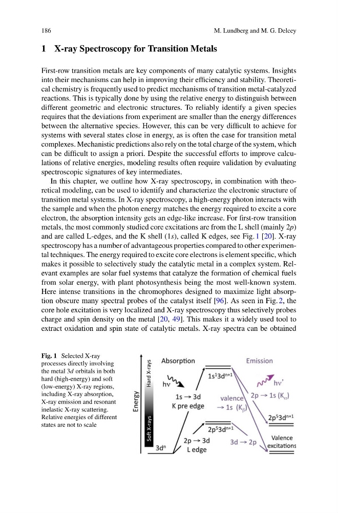
metals, the most commonly studied core excitations are from the L shell (mainly 2p)
and are called L-edges, and the K shell (1s), called K edges, see Fig. 1 [20]. X-ray
spectroscopy has a number of advantageous properties compared to other experimental techniques. The energy required to excite core electrons is element specific, which
makes it possible to selectively study the catalytic metal in a complex system. Relevant examples are solar fuel systems that catalyze the formation of chemical fuels
from solar energy, with plant photosynthesis being the most well-known system.
Here intense transitions in the chromophores designed to maximize light absorption obscure many spectral probes of the catalyst itself [96]. As seen in Fig. 2, the
core hole excitation is very localized and X-ray spectroscopy thus selectively probes
charge and spin density on the metal [20, 49]. This makes it a widely used tool to
extract oxidation and spin state of catalytic metals. X-ray spectra can be obtained
Fig. 1 Selected X-ray
processes directly involving
the metal 3d orbitals in both
hard (high-energy) and soft
(low-energy) X-ray regions,
including X-ray absorption,
X-ray emission and resonant
inelastic X-ray scattering.
Relative energies of different
states are not to scale
M. Lundberg and M. G. Delcey
1 X-ray Spectroscopy for Transition Metals
First-row transition metals are key components of many catalytic systems. Insights
into their mechanisms can help in improving their efficiency and stability. Theoretical chemistry is frequently used to predict mechanisms of transition metal-catalyzed
reactions. This is typically done by using the relative energy to distinguish between
different geometric and electronic structures. To reliably identify a given species
requires that the deviations from experiment are smaller than the energy differences
between the alternative species. However, this can be very difficult to achieve for
systems with several states close in energy, as is often the case for transition metal
complexes. Mechanistic predictions also rely on the total charge of the system, which
can be difficult to assign a priori. Despite the successful efforts to improve calculations of relative energies, modeling results often require validation by evaluating
spectroscopic signatures of key intermediates.
In this chapter, we outline how X-ray spectroscopy, in combination with theoretical modeling, can be used to identify and characterize the electronic structure of
transition metal systems. In X-ray spectroscopy, a high-energy photon interacts with
the sample and when the photon energy matches the energy required to excite a core
electron, the absorption intensity gets an edge-like increase. For first-row transition
metals, the most commonly studied core excitations are from the L shell (mainly 2p)
and are called L-edges, and the K shell (1s), called K edges, see Fig. 1 [20]. X-ray
spectroscopy has a number of advantageous properties compared to other experimental techniques. The energy required to excite core electrons is element specific, which
makes it possible to selectively study the catalytic metal in a complex system. Relevant examples are solar fuel systems that catalyze the formation of chemical fuels
from solar energy, with plant photosynthesis being the most well-known system.
Here intense transitions in the chromophores designed to maximize light absorption obscure many spectral probes of the catalyst itself [96]. As seen in Fig. 2, the
core hole excitation is very localized and X-ray spectroscopy thus selectively probes
charge and spin density on the metal [20, 49]. This makes it a widely used tool to
extract oxidation and spin state of catalytic metals. X-ray spectra can be obtained
Fig. 1 Selected X-ray
processes directly involving
the metal 3d orbitals in both
hard (high-energy) and soft
(low-energy) X-ray regions,
including X-ray absorption,
X-ray emission and resonant
inelastic X-ray scattering.
Relative energies of different
states are not to scale