

Let us now consider the interaction between a ligand and a protein active site.
The low-gradient isosurface for a tetracycline inhibitor bound to the tetR protein in
Fig. 18.7 shows a complex web of non-covalent interactions between the ligand
and the active site. When analyzing non-covalent interactions in protein-ligand
complexes, it is usually assumed that these interactions are due to a specific contact
between two atoms [44]. However, it is clearly seen in Fig. 18.7 that this
assumption is only partly correct. Hydrogen bonds, such as those between the
tetracycline amine groups and two water molecules (atoms shown in orange), are
directional and specific. Conversely, van der Waals, dipole-dipole, and hydrophobic
interactions, such as those between the tetracycline and the Leu61, Val91, Ile136,
and Val166 residues (atoms shown in yellow), are not atom-specific and occupy
broader regions in space. The figure reveals some steric clashes (orange and red
regions of the isosurface) that must be offset by stronger, attractive interactions to
give binding in this crystal structure. A ligand “fits” the geometry of the active site,
and the interaction energy between the ligand and protein is comprised of many
small contributions. When trying to design a new ligand to fit a specific active site,
one should consider all such interactions.
18.7 New Bonds: Do We Really Need to Name Them All?
Hydrogen bonding was postulated in the early twentieth century based on the
stunning macroscopic differences between the first and second row hydrides, i.e.
water is a high boiling liquid without, which there would be no life, and hydrogen
sulphide is a stinking gas, under ambient conditions [1].
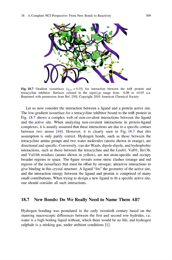
Fig. 18.7 Gradient isosurfaces (s pro = 0.35) for interaction between the tetR protein and
tetracycline inhibitor. Surfaces colored in the sign(λ 2 )ρ range from −0.06 to +0.05 a.u.
Reprinted with permission from Ref. [50]. Copyright 2010 American Chemical Society
18 A Complete NCI Perspective: From New Bonds to Reactivity
509
The low-gradient isosurface for a tetracycline inhibitor bound to the tetR protein in
Fig. 18.7 shows a complex web of non-covalent interactions between the ligand
and the active site. When analyzing non-covalent interactions in protein-ligand
complexes, it is usually assumed that these interactions are due to a specific contact
between two atoms [44]. However, it is clearly seen in Fig. 18.7 that this
assumption is only partly correct. Hydrogen bonds, such as those between the
tetracycline amine groups and two water molecules (atoms shown in orange), are
directional and specific. Conversely, van der Waals, dipole-dipole, and hydrophobic
interactions, such as those between the tetracycline and the Leu61, Val91, Ile136,
and Val166 residues (atoms shown in yellow), are not atom-specific and occupy
broader regions in space. The figure reveals some steric clashes (orange and red
regions of the isosurface) that must be offset by stronger, attractive interactions to
give binding in this crystal structure. A ligand “fits” the geometry of the active site,
and the interaction energy between the ligand and protein is comprised of many
small contributions. When trying to design a new ligand to fit a specific active site,
one should consider all such interactions.
18.7 New Bonds: Do We Really Need to Name Them All?
Hydrogen bonding was postulated in the early twentieth century based on the
stunning macroscopic differences between the first and second row hydrides, i.e.
water is a high boiling liquid without, which there would be no life, and hydrogen
sulphide is a stinking gas, under ambient conditions [1].
Fig. 18.7 Gradient isosurfaces (s pro = 0.35) for interaction between the tetR protein and
tetracycline inhibitor. Surfaces colored in the sign(λ 2 )ρ range from −0.06 to +0.05 a.u.
Reprinted with permission from Ref. [50]. Copyright 2010 American Chemical Society
18 A Complete NCI Perspective: From New Bonds to Reactivity
509