

33
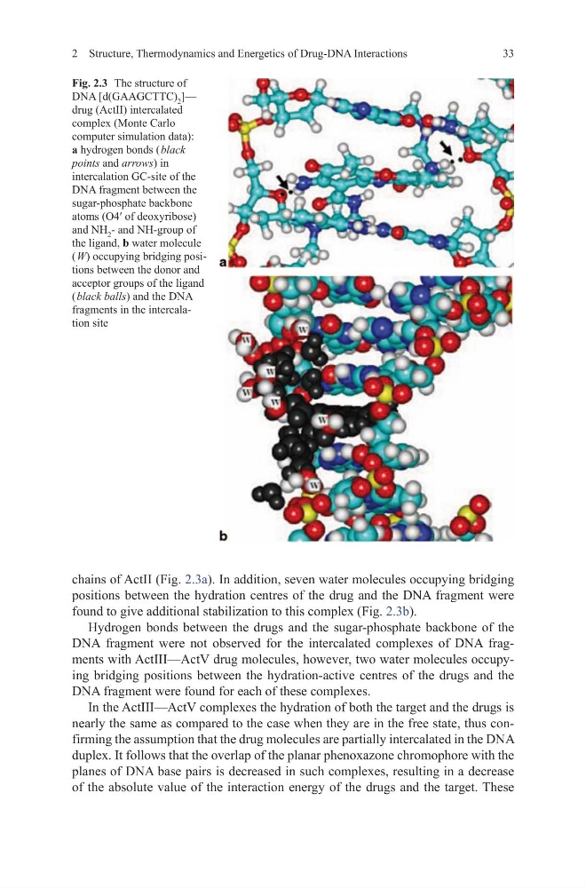
chains of ActII (Fig. 2.3a). In addition, seven water molecules occupying bridging
positions between the hydration centres of the drug and the DNA fragment were
found to give additional stabilization to this complex (Fig. 2.3b).
Hydrogen bonds between the drugs and the sugar-phosphate backbone of the
DNA fragment were not observed for the intercalated complexes of DNA fragments with ActIII—ActV drug molecules, however, two water molecules occupying bridging positions between the hydration-active centres of the drugs and the
DNA fragment were found for each of these complexes.
In the ActIII—ActV complexes the hydration of both the target and the drugs is
nearly the same as compared to the case when they are in the free state, thus confirming the assumption that the drug molecules are partially intercalated in the DNA
duplex. It follows that the overlap of the planar phenoxazone chromophore with the
planes of DNA base pairs is decreased in such complexes, resulting in a decrease
of the absolute value of the interaction energy of the drugs and the target. These
Fig. 2.3 The structure of
DNA [d(GAAGCTTC) 2 ]—
drug (ActII) intercalated
complex (Monte Carlo
computer simulation data):
а hydrogen bonds ( black
points and arrows) in
intercalation GC-site of the
DNA fragment between the
sugar-phosphate backbone
atoms (O4′ of deoxyribose)
and NH 2 - and NH-group of
the ligand, b water molecule
( W) occupying bridging positions between the donor and
acceptor groups of the ligand
( black balls) and the DNA
fragments in the intercalation site
2 Structure, Thermodynamics and Energetics of Drug-DNA Interactions
chains of ActII (Fig. 2.3a). In addition, seven water molecules occupying bridging
positions between the hydration centres of the drug and the DNA fragment were
found to give additional stabilization to this complex (Fig. 2.3b).
Hydrogen bonds between the drugs and the sugar-phosphate backbone of the
DNA fragment were not observed for the intercalated complexes of DNA fragments with ActIII—ActV drug molecules, however, two water molecules occupying bridging positions between the hydration-active centres of the drugs and the
DNA fragment were found for each of these complexes.
In the ActIII—ActV complexes the hydration of both the target and the drugs is
nearly the same as compared to the case when they are in the free state, thus confirming the assumption that the drug molecules are partially intercalated in the DNA
duplex. It follows that the overlap of the planar phenoxazone chromophore with the
planes of DNA base pairs is decreased in such complexes, resulting in a decrease
of the absolute value of the interaction energy of the drugs and the target. These
Fig. 2.3 The structure of
DNA [d(GAAGCTTC) 2 ]—
drug (ActII) intercalated
complex (Monte Carlo
computer simulation data):
а hydrogen bonds ( black
points and arrows) in
intercalation GC-site of the
DNA fragment between the
sugar-phosphate backbone
atoms (O4′ of deoxyribose)
and NH 2 - and NH-group of
the ligand, b water molecule
( W) occupying bridging positions between the donor and
acceptor groups of the ligand
( black balls) and the DNA
fragments in the intercalation site
2 Structure, Thermodynamics and Energetics of Drug-DNA Interactions