

282
D. Toczek et al.
shift of its anomeric proton. This experiment indicates the formation of complex
formation with the α-d-allopyranose only and as a consequence the appropriate
equilibrium shift. It is worth to note that the effect after the addition of monovalent
sodium ion is small due to weak complexing ability of sodium. The NMR studies
also revealed that spectra of α-furanose but not β-furanose was changed indicating that the cis–cis arrangement of five membered α-furanose ring also favors the
complex formation. The fact that only α-d-allopyranose and α-d-allofuranose form
complexes with calcium and β forms was not independently confirmed by electrophoresis experiments [17].
In the case of 5-o-methyl-d-ribose [17] α-furanose isomer consist of 33 % and
β-furanose 67 % in the solution. After the addition of calcium chloride the proportion was changed to 70:30 respectively. It is due to the possibility of α-furanose to
adopt conformation in which hydroxyl groups O1, O2, O3 are quasi axial, quasi
equatorial, quasi axial which is close to typical arrangement ax–eq–ax.
As expected, α-d-ribopyranose and α-d-ribofuranose [41] signals in NMR are
shifted downfield due to the addition of calcium chloride, since they have acquired
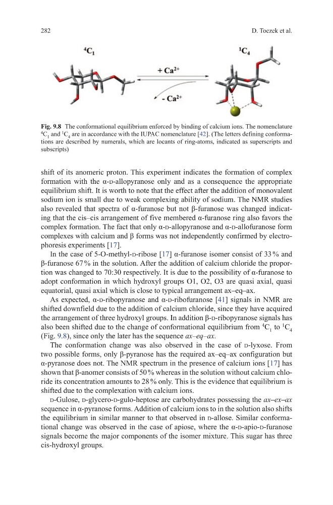
the arrangement of three hydroxyl groups. In addition β-d-ribopyranose signals has
also been shifted due to the change of conformational equilibrium from
4
C 1 to
1
C 4
(Fig. 9.8), since only the later has the sequence ax–eq–ax.
The conformation change was also observed in the case of d-lyxose. From
two possible forms, only β-pyranose has the required ax–eq–ax configuration but
α-pyranose does not. The NMR spectrum in the presence of calcium ions [17] has
shown that β-anomer consists of 50 % whereas in the solution without calcium chloride its concentration amounts to 28 % only. This is the evidence that equilibrium is
shifted due to the complexation with calcium ions.
d-Gulose, d-glycero-d-gulo-heptose are carbohydrates possessing the ax–ex–ax
sequence in α-pyranose forms. Addition of calcium ions to in the solution also shifts
the equilibrium in similar manner to that observed in d-allose. Similar conformational change was observed in the case of apiose, where the α-d-apio-d-furanose
signals become the major components of the isomer mixture. This sugar has three
cis-hydroxyl groups.
Fig. 9.8 The conformational equilibrium enforced by binding of calcium ions. The nomenclature
4
C 1 and
1
C 4 are in accordance with the IUPAC nomenclature [42]. (The letters defining conformations are described by numerals, which are locants of ring-atoms, indicated as superscripts and
subscripts)
D. Toczek et al.
shift of its anomeric proton. This experiment indicates the formation of complex
formation with the α-d-allopyranose only and as a consequence the appropriate
equilibrium shift. It is worth to note that the effect after the addition of monovalent
sodium ion is small due to weak complexing ability of sodium. The NMR studies
also revealed that spectra of α-furanose but not β-furanose was changed indicating that the cis–cis arrangement of five membered α-furanose ring also favors the
complex formation. The fact that only α-d-allopyranose and α-d-allofuranose form
complexes with calcium and β forms was not independently confirmed by electrophoresis experiments [17].
In the case of 5-o-methyl-d-ribose [17] α-furanose isomer consist of 33 % and
β-furanose 67 % in the solution. After the addition of calcium chloride the proportion was changed to 70:30 respectively. It is due to the possibility of α-furanose to
adopt conformation in which hydroxyl groups O1, O2, O3 are quasi axial, quasi
equatorial, quasi axial which is close to typical arrangement ax–eq–ax.
As expected, α-d-ribopyranose and α-d-ribofuranose [41] signals in NMR are
shifted downfield due to the addition of calcium chloride, since they have acquired
the arrangement of three hydroxyl groups. In addition β-d-ribopyranose signals has
also been shifted due to the change of conformational equilibrium from
4
C 1 to
1
C 4
(Fig. 9.8), since only the later has the sequence ax–eq–ax.
The conformation change was also observed in the case of d-lyxose. From
two possible forms, only β-pyranose has the required ax–eq–ax configuration but
α-pyranose does not. The NMR spectrum in the presence of calcium ions [17] has
shown that β-anomer consists of 50 % whereas in the solution without calcium chloride its concentration amounts to 28 % only. This is the evidence that equilibrium is
shifted due to the complexation with calcium ions.
d-Gulose, d-glycero-d-gulo-heptose are carbohydrates possessing the ax–ex–ax
sequence in α-pyranose forms. Addition of calcium ions to in the solution also shifts
the equilibrium in similar manner to that observed in d-allose. Similar conformational change was observed in the case of apiose, where the α-d-apio-d-furanose
signals become the major components of the isomer mixture. This sugar has three
cis-hydroxyl groups.
Fig. 9.8 The conformational equilibrium enforced by binding of calcium ions. The nomenclature
4
C 1 and
1
C 4 are in accordance with the IUPAC nomenclature [42]. (The letters defining conformations are described by numerals, which are locants of ring-atoms, indicated as superscripts and
subscripts)