

278
D. Toczek et al.
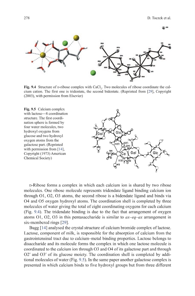
d-Ribose forms a complex in which each calcium ion is shared by two ribose
molecules. One ribose molecule represents tridendate ligand binding calcium ion
through O1, O2, O3 atoms, the second ribose is a bidendate ligand and binds via
O4 and O5 oxygen hydroxyl atoms. The coordination shell is completed by three
molecules of water giving the total of eight coordinating oxygens for each calcium
(Fig. 9.4). The tridendate binding is due to the fact that arrangement of oxygen
atoms O1, O2, O3 in this pentasaccharide is similar to ax–eq–ax arrangement in
six-membered rings [29].
Bugg [14] analysed the crystal structure of calcium bromide complex of lactose.
Lactose, component of milk, is responsible for the absorption of calcium from the
gastrointensinal tract due to calcium–metal binding properties. Lactose belongs to
disaccharide and its molecule forms the complex in which one lactose molecule is
coordinated to the calcium ion through O3 and O4 of its galactose part and through
O2′ and O3′ of its glucose moiety. The coordination shell is completed by additional molecules of water (Fig. 9.5). In the same paper another galactose complex is
presented in which calcium binds to five hydroxyl groups but from three different
Fig. 9.5 Calcium complex
with lactose—8 coordination
structure. The first coordination sphere is formed by
four water molecules, two
hydroxyl oxygens from
glucose and two hydroxyl
oxygen atoms from the
galactose part. (Reprinted
with permission from [14],
Copyright (1973) American
Chemical Society)
Fig. 9.4 Structure of d-ribose complex with CaCl 2 . Two molecules of ribose coordinate the calcium cation. The first one is tridentate, the second bidentate. (Reprinted from [29], Copyright
(2003), with permission from Elsevier)
D. Toczek et al.
d-Ribose forms a complex in which each calcium ion is shared by two ribose
molecules. One ribose molecule represents tridendate ligand binding calcium ion
through O1, O2, O3 atoms, the second ribose is a bidendate ligand and binds via
O4 and O5 oxygen hydroxyl atoms. The coordination shell is completed by three
molecules of water giving the total of eight coordinating oxygens for each calcium
(Fig. 9.4). The tridendate binding is due to the fact that arrangement of oxygen
atoms O1, O2, O3 in this pentasaccharide is similar to ax–eq–ax arrangement in
six-membered rings [29].
Bugg [14] analysed the crystal structure of calcium bromide complex of lactose.
Lactose, component of milk, is responsible for the absorption of calcium from the
gastrointensinal tract due to calcium–metal binding properties. Lactose belongs to
disaccharide and its molecule forms the complex in which one lactose molecule is
coordinated to the calcium ion through O3 and O4 of its galactose part and through
O2′ and O3′ of its glucose moiety. The coordination shell is completed by additional molecules of water (Fig. 9.5). In the same paper another galactose complex is
presented in which calcium binds to five hydroxyl groups but from three different
Fig. 9.5 Calcium complex
with lactose—8 coordination
structure. The first coordination sphere is formed by
four water molecules, two
hydroxyl oxygens from
glucose and two hydroxyl
oxygen atoms from the
galactose part. (Reprinted
with permission from [14],
Copyright (1973) American
Chemical Society)
Fig. 9.4 Structure of d-ribose complex with CaCl 2 . Two molecules of ribose coordinate the calcium cation. The first one is tridentate, the second bidentate. (Reprinted from [29], Copyright
(2003), with permission from Elsevier)