

213
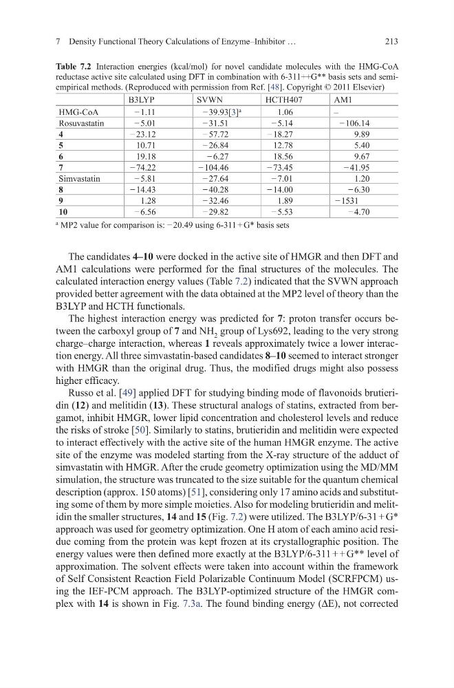
The candidates 4–10 were docked in the active site of HMGR and then DFT and
AM1 calculations were performed for the final structures of the molecules. The
calculated interaction energy values (Table 7.2) indicated that the SVWN approach
provided better agreement with the data obtained at the MP2 level of theory than the
B3LYP and HCTH functionals.
The highest interaction energy was predicted for 7: proton transfer occurs between the carboxyl group of 7 and NH 2 group of Lys692, leading to the very strong
charge–charge interaction, whereas 1 reveals approximately twice a lower interaction energy. All three simvastatin-based candidates 8–10 seemed to interact stronger
with HMGR than the original drug. Thus, the modified drugs might also possess
higher efficacy.
Russo et al. [49] applied DFT for studying binding mode of flavonoids brutieridin (12) and melitidin (13). These structural analogs of statins, extracted from bergamot, inhibit HMGR, lower lipid concentration and cholesterol levels and reduce
the risks of stroke [50]. Similarly to statins, brutieridin and melitidin were expected
to interact effectively with the active site of the human HMGR enzyme. The active
site of the enzyme was modeled starting from the X-ray structure of the adduct of
simvastatin with HMGR. After the crude geometry optimization using the MD/MM
simulation, the structure was truncated to the size suitable for the quantum chemical
description (approx. 150 atoms) [51], considering only 17 amino acids and substituting some of them by more simple moieties. Also for modeling brutieridin and melitidin the smaller structures, 14 and 15 (Fig. 7.2) were utilized. The B3LYP/6-31 + G*
approach was used for geometry optimization. One H atom of each amino acid residue coming from the protein was kept frozen at its crystallographic position. The
energy values were then defined more exactly at the B3LYP/6-311 + + G** level of
approximation. The solvent effects were taken into account within the framework
of Self Consistent Reaction Field Polarizable Continuum Model (SCRFPCM) using the IEF-PCM approach. The B3LYP-optimized structure of the HMGR complex with 14 is shown in Fig. 7.3a. The found binding energy (∆E), not corrected
Table 7.2 Interaction energies (kcal/mol) for novel candidate molecules with the HMG-CoA
reductase active site calculated using DFT in combination with 6-311++G** basis sets and semiempirical methods. (Reproduced with permission from Ref. [48]. Copyright © 2011 Elsevier)
B3LYP
SVWN
HCTH407
AM1
HMG-CoA
− 1.11
− 39.93[3]
a
1.06
–
Rosuvastatin
− 5.01
− 31.51
− 5.14
− 106.14
4
− 23.12
− 57.72
− 18.27
9.89
5
10.71
− 26.84
12.78
5.40
6
19.18
− 6.27
18.56
9.67
7
− 74.22
− 104.46
− 73.45
− 41.95
Simvastatin
− 5.81
− 27.64
− 7.01
1.20
8
− 14.43
− 40.28
− 14.00
− 6.30
9
1.28
− 32.46
1.89
− 1531
10
− 6.56
− 29.82
− 5.53
− 4.70
a
MP2 value for comparison is: − 20.49 using 6-311 + G* basis sets
7 Density Functional Theory Calculations of Enzyme–Inhibitor …
The candidates 4–10 were docked in the active site of HMGR and then DFT and
AM1 calculations were performed for the final structures of the molecules. The
calculated interaction energy values (Table 7.2) indicated that the SVWN approach
provided better agreement with the data obtained at the MP2 level of theory than the
B3LYP and HCTH functionals.
The highest interaction energy was predicted for 7: proton transfer occurs between the carboxyl group of 7 and NH 2 group of Lys692, leading to the very strong
charge–charge interaction, whereas 1 reveals approximately twice a lower interaction energy. All three simvastatin-based candidates 8–10 seemed to interact stronger
with HMGR than the original drug. Thus, the modified drugs might also possess
higher efficacy.
Russo et al. [49] applied DFT for studying binding mode of flavonoids brutieridin (12) and melitidin (13). These structural analogs of statins, extracted from bergamot, inhibit HMGR, lower lipid concentration and cholesterol levels and reduce
the risks of stroke [50]. Similarly to statins, brutieridin and melitidin were expected
to interact effectively with the active site of the human HMGR enzyme. The active
site of the enzyme was modeled starting from the X-ray structure of the adduct of
simvastatin with HMGR. After the crude geometry optimization using the MD/MM
simulation, the structure was truncated to the size suitable for the quantum chemical
description (approx. 150 atoms) [51], considering only 17 amino acids and substituting some of them by more simple moieties. Also for modeling brutieridin and melitidin the smaller structures, 14 and 15 (Fig. 7.2) were utilized. The B3LYP/6-31 + G*
approach was used for geometry optimization. One H atom of each amino acid residue coming from the protein was kept frozen at its crystallographic position. The
energy values were then defined more exactly at the B3LYP/6-311 + + G** level of
approximation. The solvent effects were taken into account within the framework
of Self Consistent Reaction Field Polarizable Continuum Model (SCRFPCM) using the IEF-PCM approach. The B3LYP-optimized structure of the HMGR complex with 14 is shown in Fig. 7.3a. The found binding energy (∆E), not corrected
Table 7.2 Interaction energies (kcal/mol) for novel candidate molecules with the HMG-CoA
reductase active site calculated using DFT in combination with 6-311++G** basis sets and semiempirical methods. (Reproduced with permission from Ref. [48]. Copyright © 2011 Elsevier)
B3LYP
SVWN
HCTH407
AM1
HMG-CoA
− 1.11
− 39.93[3]
a
1.06
–
Rosuvastatin
− 5.01
− 31.51
− 5.14
− 106.14
4
− 23.12
− 57.72
− 18.27
9.89
5
10.71
− 26.84
12.78
5.40
6
19.18
− 6.27
18.56
9.67
7
− 74.22
− 104.46
− 73.45
− 41.95
Simvastatin
− 5.81
− 27.64
− 7.01
1.20
8
− 14.43
− 40.28
− 14.00
− 6.30
9
1.28
− 32.46
1.89
− 1531
10
− 6.56
− 29.82
− 5.53
− 4.70
a
MP2 value for comparison is: − 20.49 using 6-311 + G* basis sets
7 Density Functional Theory Calculations of Enzyme–Inhibitor …