

117
Appropriate binding sites are localized on the DNA template binding interface sufficiently close to each other and can contain shared amino acids. The binding of any
of the above-considered low-weight molecular compounds directly prevents interaction with ssDNA and defines the competitive character of polymerase inhibition.
The computational modeling of protein–ligand interactions was performed also
for compounds inhibiting other DNA-dependent DNA polymerases. So, structural
insights of actions were investigated in silico for high specific pol λ inhibitors—
petasiphenol [95] as well as curcumin and its derivate monoacetylcurcumin [86].
The petasiphenol is a natural phenolic compound produced from a higher plant,
a Japanese vegetable ( Petasites japonicus) that was originally found to be a bioantimutagen in UV-induced mutagenic Escherichia coli WP2 B/r Trp-isolated from
the same plant [123]. It was established experimentally that petasiphenol binds to
N-terminal BRCT domain of pol λ with IC 50 of 7.6 µM and does not bind to the Cterminal catalytic domain including the pol β-like core of pol λ [95].
Spatial structure of pol λ BRCT domain was reconstructed via homology modeling using molecular modeling software Insight II (module Homology) (Accelrys
Inc., San Diego, CA). The spatial structure of human XRCC1 (PDB accession code
is 1CDZ [124]) was used as a template for the modeling. Molecular docking of
petasiphenol molecule into pol λ BRCT domain and the further binding evaluation were performed according to the procedures described above for solanapyrone
A. It was revealed that the N-terminal BRCT domain of pol λ (residues 36–132)
consists of three α-helices and four β-sheets. The petasiphenol-binding region in
the BRCT domain of pol λ is assumed to consist of the two loops (residues 74–81
and 84–107) between the β-sheet (residues 82–83) and includes amino acids Gln76,
Ile83, Asp90, Glu92, Arg93 Ala94 Leu95, Arg96, Leu98, Arg99, Leu100, Gln102,
Leu103 and Pro104 [95].
The hydroxyl and ketone groups of petasiphenol may show a preference for binding to the hydrophilic residue of Gln76, Arg93, and Arg99, and, on the other hand,
the benzene groups may be absorbed to the hydrophobic amino acids in the loops.
The binding energies between NH 2 of Gln76, NH 2
+
of Arg93, or NH 2
+
of Arg99 and
the hydrophilic groups in petasiphenol were − 9.400, − 3.652 and − 4.642 kcal/mol
respectively, and the binding force consisted of the Coulomb force (− 7.904, − 2.220
4 DNA Dependent DNA Polymerases as Targets for Low-Weight …
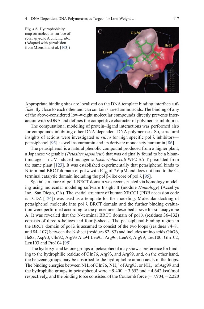
Fig. 4.6 Hydrophobicity
map on molecular surface of
solanapyrone A binding site.
(Adapted with permission
from Mizushina et al. [103])
Appropriate binding sites are localized on the DNA template binding interface sufficiently close to each other and can contain shared amino acids. The binding of any
of the above-considered low-weight molecular compounds directly prevents interaction with ssDNA and defines the competitive character of polymerase inhibition.
The computational modeling of protein–ligand interactions was performed also
for compounds inhibiting other DNA-dependent DNA polymerases. So, structural
insights of actions were investigated in silico for high specific pol λ inhibitors—
petasiphenol [95] as well as curcumin and its derivate monoacetylcurcumin [86].
The petasiphenol is a natural phenolic compound produced from a higher plant,
a Japanese vegetable ( Petasites japonicus) that was originally found to be a bioantimutagen in UV-induced mutagenic Escherichia coli WP2 B/r Trp-isolated from
the same plant [123]. It was established experimentally that petasiphenol binds to
N-terminal BRCT domain of pol λ with IC 50 of 7.6 µM and does not bind to the Cterminal catalytic domain including the pol β-like core of pol λ [95].
Spatial structure of pol λ BRCT domain was reconstructed via homology modeling using molecular modeling software Insight II (module Homology) (Accelrys
Inc., San Diego, CA). The spatial structure of human XRCC1 (PDB accession code
is 1CDZ [124]) was used as a template for the modeling. Molecular docking of
petasiphenol molecule into pol λ BRCT domain and the further binding evaluation were performed according to the procedures described above for solanapyrone
A. It was revealed that the N-terminal BRCT domain of pol λ (residues 36–132)
consists of three α-helices and four β-sheets. The petasiphenol-binding region in
the BRCT domain of pol λ is assumed to consist of the two loops (residues 74–81
and 84–107) between the β-sheet (residues 82–83) and includes amino acids Gln76,
Ile83, Asp90, Glu92, Arg93 Ala94 Leu95, Arg96, Leu98, Arg99, Leu100, Gln102,
Leu103 and Pro104 [95].
The hydroxyl and ketone groups of petasiphenol may show a preference for binding to the hydrophilic residue of Gln76, Arg93, and Arg99, and, on the other hand,
the benzene groups may be absorbed to the hydrophobic amino acids in the loops.
The binding energies between NH 2 of Gln76, NH 2
+
of Arg93, or NH 2
+
of Arg99 and
the hydrophilic groups in petasiphenol were − 9.400, − 3.652 and − 4.642 kcal/mol
respectively, and the binding force consisted of the Coulomb force (− 7.904, − 2.220
4 DNA Dependent DNA Polymerases as Targets for Low-Weight …
Fig. 4.6 Hydrophobicity
map on molecular surface of
solanapyrone A binding site.
(Adapted with permission
from Mizushina et al. [103])