

116
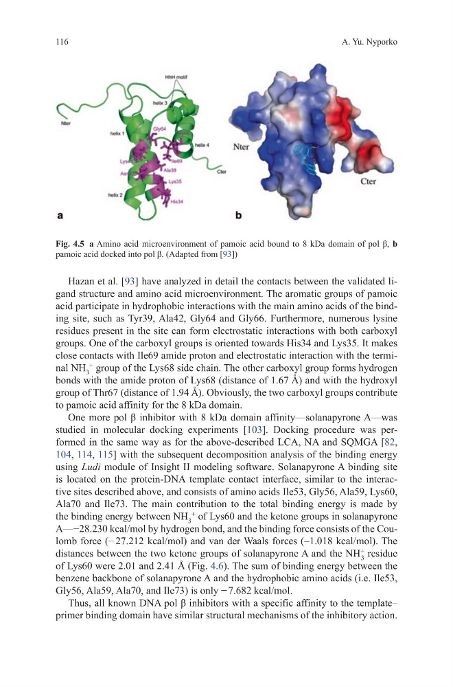
Hazan et al. [93] have analyzed in detail the contacts between the validated ligand structure and amino acid microenvironment. The aromatic groups of pamoic
acid participate in hydrophobic interactions with the main amino acids of the binding site, such as Tyr39, Ala42, Gly64 and Gly66. Furthermore, numerous lysine
residues present in the site can form electrostatic interactions with both carboxyl
groups. One of the carboxyl groups is oriented towards His34 and Lys35. It makes
close contacts with Ile69 amide proton and electrostatic interaction with the terminal NH 3
+
group of the Lys68 side chain. The other carboxyl group forms hydrogen
bonds with the amide proton of Lys68 (distance of 1.67 A ˚ ) and with the hydroxyl
group of Thr67 (distance of 1.94 A ˚ ). Obviously, the two carboxyl groups contribute
to pamoic acid affinity for the 8 kDa domain.
One more pol β inhibitor with 8 kDa domain affinity—solanapyrone A—was
studied in molecular docking experiments [103]. Docking procedure was performed in the same way as for the above-described LCA, NA and SQMGA [82,
104, 114, 115] with the subsequent decomposition analysis of the binding energy
using Ludi module of Insight II modeling software. Solanapyrone A binding site
is located on the protein-DNA template contact interface, similar to the interactive sites described above, and consists of amino acids Ile53, Gly56, Ala59, Lys60,
Ala70 and Ile73. The main contribution to the total binding energy is made by
the binding energy between NH 3
+
of Lys60 and the ketone groups in solanapyrone
A—−28.230 kcal/mol by hydrogen bond, and the binding force consists of the Coulomb force (− 27.212 kcal/mol) and van der Waals forces (–1.018 kcal/mol). The
distances between the two ketone groups of solanapyrone A and the NH
+
3
residue
of Lys60 were 2.01 and 2.41 Å (Fig. 4.6). The sum of binding energy between the
benzene backbone of solanapyrone A and the hydrophobic amino acids (i.e. Ile53,
Gly56, Ala59, Ala70, and Ile73) is only − 7.682 kcal/mol.
Thus, all known DNA pol β inhibitors with a specific affinity to the template–
primer binding domain have similar structural mechanisms of the inhibitory action.
Fig. 4.5 a Amino acid microenvironment of pamoic acid bound to 8 kDa domain of pol β, b
pamoic acid docked into pol β. (Adapted from [93])
A. Yu. Nyporko
Hazan et al. [93] have analyzed in detail the contacts between the validated ligand structure and amino acid microenvironment. The aromatic groups of pamoic
acid participate in hydrophobic interactions with the main amino acids of the binding site, such as Tyr39, Ala42, Gly64 and Gly66. Furthermore, numerous lysine
residues present in the site can form electrostatic interactions with both carboxyl
groups. One of the carboxyl groups is oriented towards His34 and Lys35. It makes
close contacts with Ile69 amide proton and electrostatic interaction with the terminal NH 3
+
group of the Lys68 side chain. The other carboxyl group forms hydrogen
bonds with the amide proton of Lys68 (distance of 1.67 A ˚ ) and with the hydroxyl
group of Thr67 (distance of 1.94 A ˚ ). Obviously, the two carboxyl groups contribute
to pamoic acid affinity for the 8 kDa domain.
One more pol β inhibitor with 8 kDa domain affinity—solanapyrone A—was
studied in molecular docking experiments [103]. Docking procedure was performed in the same way as for the above-described LCA, NA and SQMGA [82,
104, 114, 115] with the subsequent decomposition analysis of the binding energy
using Ludi module of Insight II modeling software. Solanapyrone A binding site
is located on the protein-DNA template contact interface, similar to the interactive sites described above, and consists of amino acids Ile53, Gly56, Ala59, Lys60,
Ala70 and Ile73. The main contribution to the total binding energy is made by
the binding energy between NH 3
+
of Lys60 and the ketone groups in solanapyrone
A—−28.230 kcal/mol by hydrogen bond, and the binding force consists of the Coulomb force (− 27.212 kcal/mol) and van der Waals forces (–1.018 kcal/mol). The
distances between the two ketone groups of solanapyrone A and the NH
+
3
residue
of Lys60 were 2.01 and 2.41 Å (Fig. 4.6). The sum of binding energy between the
benzene backbone of solanapyrone A and the hydrophobic amino acids (i.e. Ile53,
Gly56, Ala59, Ala70, and Ile73) is only − 7.682 kcal/mol.
Thus, all known DNA pol β inhibitors with a specific affinity to the template–
primer binding domain have similar structural mechanisms of the inhibitory action.
Fig. 4.5 a Amino acid microenvironment of pamoic acid bound to 8 kDa domain of pol β, b
pamoic acid docked into pol β. (Adapted from [93])
A. Yu. Nyporko