

2. Count cells using hemocytometer to determine the cell density.
3. Centrifuging cell samples at 135 Â g for 5 min and resuspend
cells in GIBCO Opti-MEM at a concentration of 0.5 Â 10
7
cells/mL.
4. Preset the electroporation parameters on an electroporator
(ECM 830, Harvard Apparatus).
5. Mix 20 μL of cell suspension with 1 μg Plasmid DNA (pGFP or
pLuc) and load into the MAE chamber with micropillar array
electrode surface facing upwards (the positive electrode). When
cotransfecting plasmid DNA and corresponding siRNA probes,
5 or 30 pM siRNA is added in the cell suspension and gently
mixed with pipette prior to the sample loading.
6. Take the gold-coated plain electrode (as the negative electrode
with the gold-coating surface face down) and place on the cell
solution drop in the electroporation chamber. Allow its own
weight squeeze to allow the cell solution to spread in the cell
loading chamber. Slowly push from the other side of the plain
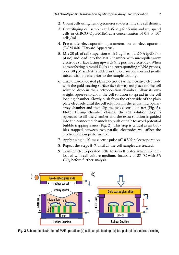
plate electrode until the cell solution fills the entire micropillararray chamber and then clip the two electrode plates (Fig. 3).
Note: During chamber closing, the cell solution drop is
squeezed to fill the chamber and the extra solution is guided
into the connected channels to push out air to avoid potential
bubble trapping issues (Fig. 2). This step is critical as air bubbles trapped between two parallel electrodes will affect the
electroporation performance.
7. Apply a single, 10-ms electric pulse of 10 V for electroporation.
8. Repeat the steps 5–7 until all the cell samples are treated.
9. Transfer electroporated cells to 6-well plates which are preloaded with cell culture medium. Incubate at 37
C with 5%
CO 2 before further analysis.
(b)
(a)
rubber gasket
Cell
Rubber Cushion
Gold coated glass slide
epoxy spacer
Silicon
micropillars
10m
m
4mm
Rubber Cushion
Gold coated glass slide
Silicon
160mm
Fig. 3 Schematic illustration of MAE operation: (a) cell sample loading; (b) top plain plate electrode closing
Cell Size-Specific Transfection by Micropillar Array Electroporation
7
3. Centrifuging cell samples at 135 Â g for 5 min and resuspend
cells in GIBCO Opti-MEM at a concentration of 0.5 Â 10
7
cells/mL.
4. Preset the electroporation parameters on an electroporator
(ECM 830, Harvard Apparatus).
5. Mix 20 μL of cell suspension with 1 μg Plasmid DNA (pGFP or
pLuc) and load into the MAE chamber with micropillar array
electrode surface facing upwards (the positive electrode). When
cotransfecting plasmid DNA and corresponding siRNA probes,
5 or 30 pM siRNA is added in the cell suspension and gently
mixed with pipette prior to the sample loading.
6. Take the gold-coated plain electrode (as the negative electrode
with the gold-coating surface face down) and place on the cell
solution drop in the electroporation chamber. Allow its own
weight squeeze to allow the cell solution to spread in the cell
loading chamber. Slowly push from the other side of the plain
plate electrode until the cell solution fills the entire micropillararray chamber and then clip the two electrode plates (Fig. 3).
Note: During chamber closing, the cell solution drop is
squeezed to fill the chamber and the extra solution is guided
into the connected channels to push out air to avoid potential
bubble trapping issues (Fig. 2). This step is critical as air bubbles trapped between two parallel electrodes will affect the
electroporation performance.
7. Apply a single, 10-ms electric pulse of 10 V for electroporation.
8. Repeat the steps 5–7 until all the cell samples are treated.
9. Transfer electroporated cells to 6-well plates which are preloaded with cell culture medium. Incubate at 37
C with 5%
CO 2 before further analysis.
(b)
(a)
rubber gasket
Cell
Rubber Cushion
Gold coated glass slide
epoxy spacer
Silicon
micropillars
10m
m
4mm
Rubber Cushion
Gold coated glass slide
Silicon
160mm
Fig. 3 Schematic illustration of MAE operation: (a) cell sample loading; (b) top plain plate electrode closing
Cell Size-Specific Transfection by Micropillar Array Electroporation
7