

32
2 Experimental and Theoretical Considerations
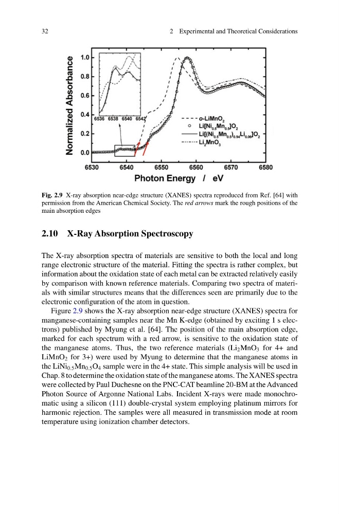
Fig. 2.9 X-ray absorption near-edge structure (XANES) spectra reproduced from Ref. [64] with
permission from the American Chemical Society. The red arrows mark the rough positions of the
main absorption edges
2.10 X-Ray Absorption Spectroscopy
The X-ray absorption spectra of materials are sensitive to both the local and long
range electronic structure of the material. Fitting the spectra is rather complex, but
information about the oxidation state of each metal can be extracted relatively easily
by comparison with known reference materials. Comparing two spectra of materials with similar structures means that the differences seen are primarily due to the
electronic configuration of the atom in question.
Figure 2.9 shows the X-ray absorption near-edge structure (XANES) spectra for
manganese-containing samples near the Mn K-edge (obtained by exciting 1 s electrons) published by Myung et al. [64]. The position of the main absorption edge,
marked for each spectrum with a red arrow, is sensitive to the oxidation state of
the manganese atoms. Thus, the two reference materials (Li 2 MnO 3 for 4+ and
LiMnO 2 for 3+) were used by Myung to determine that the manganese atoms in
the LiNi 0.5 Mn 0.5 O 4 sample were in the 4+ state. This simple analysis will be used in
Chap. 8 to determine the oxidation state of the manganese atoms. The XANES spectra
were collected by Paul Duchesne on the PNC-CAT beamline 20-BM at the Advanced
Photon Source of Argonne National Labs. Incident X-rays were made monochromatic using a silicon (111) double-crystal system employing platinum mirrors for
harmonic rejection. The samples were all measured in transmission mode at room
temperature using ionization chamber detectors.
2 Experimental and Theoretical Considerations
Fig. 2.9 X-ray absorption near-edge structure (XANES) spectra reproduced from Ref. [64] with
permission from the American Chemical Society. The red arrows mark the rough positions of the
main absorption edges
2.10 X-Ray Absorption Spectroscopy
The X-ray absorption spectra of materials are sensitive to both the local and long
range electronic structure of the material. Fitting the spectra is rather complex, but
information about the oxidation state of each metal can be extracted relatively easily
by comparison with known reference materials. Comparing two spectra of materials with similar structures means that the differences seen are primarily due to the
electronic configuration of the atom in question.
Figure 2.9 shows the X-ray absorption near-edge structure (XANES) spectra for
manganese-containing samples near the Mn K-edge (obtained by exciting 1 s electrons) published by Myung et al. [64]. The position of the main absorption edge,
marked for each spectrum with a red arrow, is sensitive to the oxidation state of
the manganese atoms. Thus, the two reference materials (Li 2 MnO 3 for 4+ and
LiMnO 2 for 3+) were used by Myung to determine that the manganese atoms in
the LiNi 0.5 Mn 0.5 O 4 sample were in the 4+ state. This simple analysis will be used in
Chap. 8 to determine the oxidation state of the manganese atoms. The XANES spectra
were collected by Paul Duchesne on the PNC-CAT beamline 20-BM at the Advanced
Photon Source of Argonne National Labs. Incident X-rays were made monochromatic using a silicon (111) double-crystal system employing platinum mirrors for
harmonic rejection. The samples were all measured in transmission mode at room
temperature using ionization chamber detectors.