

8 Ultrafast Ionization and Fragmentation: From Small Molecules
185
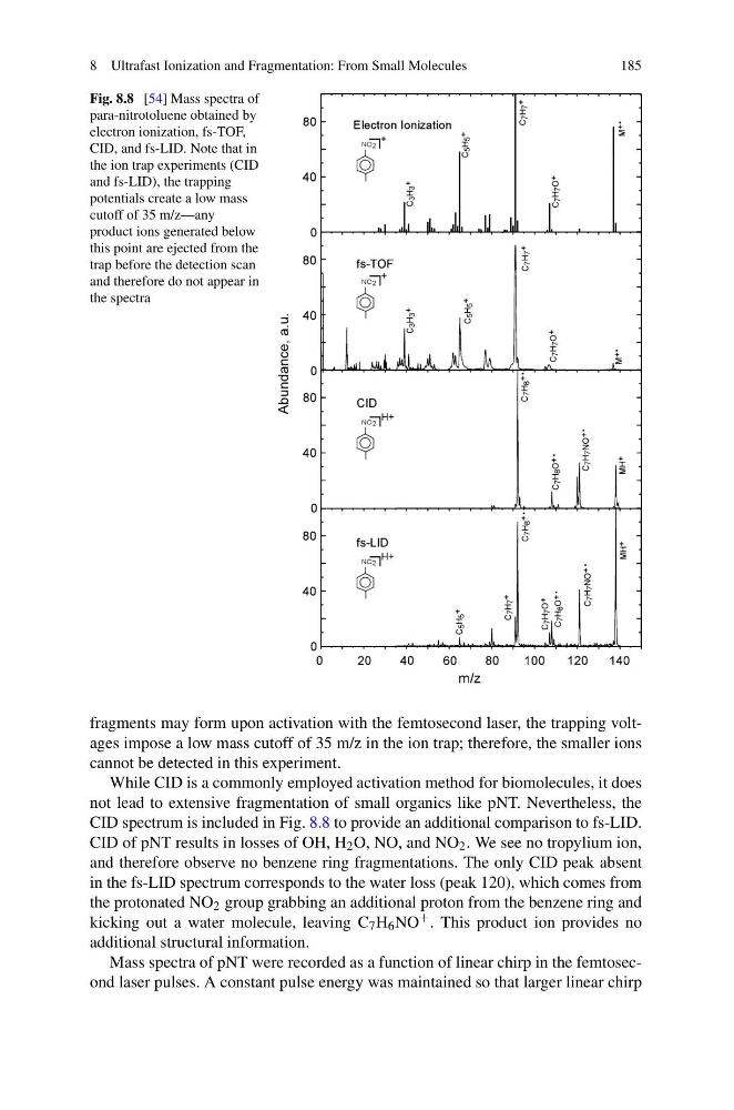
Fig. 8.8 [54] Mass spectra of
para-nitrotoluene obtained by
electron ionization, fs-TOF,
CID, and fs-LID. Note that in
the ion trap experiments (CID
and fs-LID), the trapping
potentials create a low mass
cutoff of 35 m/z—any
product ions generated below
this point are ejected from the
trap before the detection scan
and therefore do not appear in
the spectra
fragments may form upon activation with the femtosecond laser, the trapping voltages impose a low mass cutoff of 35 m/z in the ion trap; therefore, the smaller ions
cannot be detected in this experiment.
While CID is a commonly employed activation method for biomolecules, it does
not lead to extensive fragmentation of small organics like pNT. Nevertheless, the
CID spectrum is included in Fig. 8.8 to provide an additional comparison to fs-LID.
CID of pNT results in losses of OH, H 2 O, NO, and NO 2 . We see no tropylium ion,
and therefore observe no benzene ring fragmentations. The only CID peak absent
in the fs-LID spectrum corresponds to the water loss (peak 120), which comes from
the protonated NO 2 group grabbing an additional proton from the benzene ring and
kicking out a water molecule, leaving C 7 H 6 NO
+ . This product ion provides no
additional structural information.
Mass spectra of pNT were recorded as a function of linear chirp in the femtosecond laser pulses. A constant pulse energy was maintained so that larger linear chirp
185
Fig. 8.8 [54] Mass spectra of
para-nitrotoluene obtained by
electron ionization, fs-TOF,
CID, and fs-LID. Note that in
the ion trap experiments (CID
and fs-LID), the trapping
potentials create a low mass
cutoff of 35 m/z—any
product ions generated below
this point are ejected from the
trap before the detection scan
and therefore do not appear in
the spectra
fragments may form upon activation with the femtosecond laser, the trapping voltages impose a low mass cutoff of 35 m/z in the ion trap; therefore, the smaller ions
cannot be detected in this experiment.
While CID is a commonly employed activation method for biomolecules, it does
not lead to extensive fragmentation of small organics like pNT. Nevertheless, the
CID spectrum is included in Fig. 8.8 to provide an additional comparison to fs-LID.
CID of pNT results in losses of OH, H 2 O, NO, and NO 2 . We see no tropylium ion,
and therefore observe no benzene ring fragmentations. The only CID peak absent
in the fs-LID spectrum corresponds to the water loss (peak 120), which comes from
the protonated NO 2 group grabbing an additional proton from the benzene ring and
kicking out a water molecule, leaving C 7 H 6 NO
+ . This product ion provides no
additional structural information.
Mass spectra of pNT were recorded as a function of linear chirp in the femtosecond laser pulses. A constant pulse energy was maintained so that larger linear chirp