

6 Biomolecules, Photostability and 1 πσ ∗ States
125
microchannel plate (MCP) assembly, which possesses very high ion detection sensitivity (> 80 % [45]).
With regards to probing the timescales for H-atom elimination, a fs pump pulse
photoexcites the molecules of interest, defining the zero of time (t = 0) for the
experiment, while a time delayed fs probe pulse, centered at 243 nm, ionizes any
H-atom photoproducts through (2 + 1) resonance enhanced multiphoton ionization
(REMPI), generating H + . Both fs pump and probe pulses are typically derived from
optical parametric amplifiers (OPAs) [46], commonly seeded by the output of a commercial ultrafast Ti:sapphire laser system. All ions, including H + , are then analyzed
using TOF-MS. By recording a mass spectrum at a series of different time delays
(t) between the fs pump and probe pulses, and integrating only the H + counts
in the mass spectrum at each t, the buildup of H + can be tracked in real time.
The generated H + signal transient can then be modeled using appropriate functions (e.g. an exponential rise function convoluted with the temporal instrument
response function—see Sect. 6.4.1) to obtain a time-constant, τ , for the appearance
of H-atoms. However, as discussed in Sect. 6.2.1, the mere detection of H-atoms is
not necessarily indicative of 1 πσ ∗ mediated dynamics. A more complete picture of
1 πσ ∗ mediated H-atom elimination can be attained using time-resolved photofragment translational spectroscopies, such as TR-VMI, which can monitor both the
ultrafast appearance timescales and KEs of any H photoproducts.
6.3.2 Time-Resolved Velocity Map Ion Imaging
Charged particle imaging techniques [47] and, in particular, velocity map ion imaging (VMI) [37], have revolutionized the field of photofragment spectroscopy and
are now essential methodologies used in gas phase molecular reaction dynamics
[48]. One of the major advantages of VMI is that it simultaneously recovers both
the recoil speed and the angular recoil trajectories (velocity vectors) of the original
3-D distribution of ionized photofragments (Newton sphere) by collecting its 2-D
projection. With respect to the schematic in Fig. 6.3, VMI is typically implemented
using a gridless Wiley-McLaren TOF electrode arrangement, which both temporally
and spatially focus ions onto a position sensitive detector (typically a pair of MCPs
coupled to a phosphor screen) placed at the terminus of a field-free flight tube. By
temporally gating the detector, it is possible to collect the 2-D projection of only
a specific photofragment mass of interest, m f , based on its known TOF, t, to the
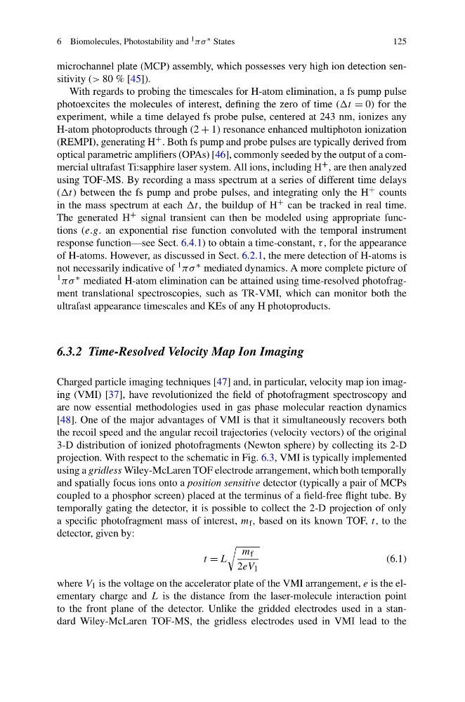
detector, given by:
t = L
m f
2eV 1
(6.1)
where V 1 is the voltage on the accelerator plate of the VMI arrangement, e is the elementary charge and L is the distance from the laser-molecule interaction point
to the front plane of the detector. Unlike the gridded electrodes used in a standard Wiley-McLaren TOF-MS, the gridless electrodes used in VMI lead to the
125
microchannel plate (MCP) assembly, which possesses very high ion detection sensitivity (> 80 % [45]).
With regards to probing the timescales for H-atom elimination, a fs pump pulse
photoexcites the molecules of interest, defining the zero of time (t = 0) for the
experiment, while a time delayed fs probe pulse, centered at 243 nm, ionizes any
H-atom photoproducts through (2 + 1) resonance enhanced multiphoton ionization
(REMPI), generating H + . Both fs pump and probe pulses are typically derived from
optical parametric amplifiers (OPAs) [46], commonly seeded by the output of a commercial ultrafast Ti:sapphire laser system. All ions, including H + , are then analyzed
using TOF-MS. By recording a mass spectrum at a series of different time delays
(t) between the fs pump and probe pulses, and integrating only the H + counts
in the mass spectrum at each t, the buildup of H + can be tracked in real time.
The generated H + signal transient can then be modeled using appropriate functions (e.g. an exponential rise function convoluted with the temporal instrument
response function—see Sect. 6.4.1) to obtain a time-constant, τ , for the appearance
of H-atoms. However, as discussed in Sect. 6.2.1, the mere detection of H-atoms is
not necessarily indicative of 1 πσ ∗ mediated dynamics. A more complete picture of
1 πσ ∗ mediated H-atom elimination can be attained using time-resolved photofragment translational spectroscopies, such as TR-VMI, which can monitor both the
ultrafast appearance timescales and KEs of any H photoproducts.
6.3.2 Time-Resolved Velocity Map Ion Imaging
Charged particle imaging techniques [47] and, in particular, velocity map ion imaging (VMI) [37], have revolutionized the field of photofragment spectroscopy and
are now essential methodologies used in gas phase molecular reaction dynamics
[48]. One of the major advantages of VMI is that it simultaneously recovers both
the recoil speed and the angular recoil trajectories (velocity vectors) of the original
3-D distribution of ionized photofragments (Newton sphere) by collecting its 2-D
projection. With respect to the schematic in Fig. 6.3, VMI is typically implemented
using a gridless Wiley-McLaren TOF electrode arrangement, which both temporally
and spatially focus ions onto a position sensitive detector (typically a pair of MCPs
coupled to a phosphor screen) placed at the terminus of a field-free flight tube. By
temporally gating the detector, it is possible to collect the 2-D projection of only
a specific photofragment mass of interest, m f , based on its known TOF, t, to the
detector, given by:
t = L
m f
2eV 1
(6.1)
where V 1 is the voltage on the accelerator plate of the VMI arrangement, e is the elementary charge and L is the distance from the laser-molecule interaction point
to the front plane of the detector. Unlike the gridded electrodes used in a standard Wiley-McLaren TOF-MS, the gridless electrodes used in VMI lead to the