

124
G.M. Roberts and V.G. Stavros
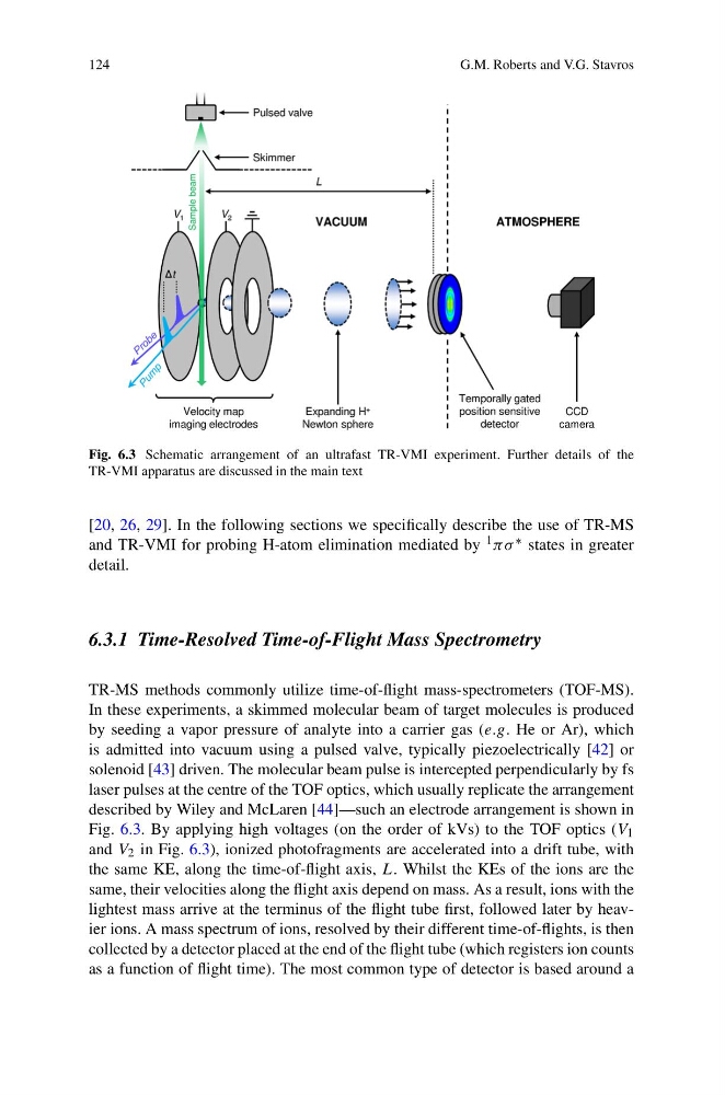
Fig. 6.3 Schematic arrangement of an ultrafast TR-VMI experiment. Further details of the
TR-VMI apparatus are discussed in the main text
[20, 26, 29]. In the following sections we specifically describe the use of TR-MS
and TR-VMI for probing H-atom elimination mediated by 1 πσ ∗ states in greater
detail.
6.3.1 Time-Resolved Time-of-Flight Mass Spectrometry
TR-MS methods commonly utilize time-of-flight mass-spectrometers (TOF-MS).
In these experiments, a skimmed molecular beam of target molecules is produced
by seeding a vapor pressure of analyte into a carrier gas (e.g. He or Ar), which
is admitted into vacuum using a pulsed valve, typically piezoelectrically [42] or
solenoid [43] driven. The molecular beam pulse is intercepted perpendicularly by fs
laser pulses at the centre of the TOF optics, which usually replicate the arrangement
described by Wiley and McLaren [44]—such an electrode arrangement is shown in
Fig. 6.3. By applying high voltages (on the order of kVs) to the TOF optics (V 1
and V 2 in Fig. 6.3), ionized photofragments are accelerated into a drift tube, with
the same KE, along the time-of-flight axis, L. Whilst the KEs of the ions are the
same, their velocities along the flight axis depend on mass. As a result, ions with the
lightest mass arrive at the terminus of the flight tube first, followed later by heavier ions. A mass spectrum of ions, resolved by their different time-of-flights, is then
collected by a detector placed at the end of the flight tube (which registers ion counts
as a function of flight time). The most common type of detector is based around a
G.M. Roberts and V.G. Stavros
Fig. 6.3 Schematic arrangement of an ultrafast TR-VMI experiment. Further details of the
TR-VMI apparatus are discussed in the main text
[20, 26, 29]. In the following sections we specifically describe the use of TR-MS
and TR-VMI for probing H-atom elimination mediated by 1 πσ ∗ states in greater
detail.
6.3.1 Time-Resolved Time-of-Flight Mass Spectrometry
TR-MS methods commonly utilize time-of-flight mass-spectrometers (TOF-MS).
In these experiments, a skimmed molecular beam of target molecules is produced
by seeding a vapor pressure of analyte into a carrier gas (e.g. He or Ar), which
is admitted into vacuum using a pulsed valve, typically piezoelectrically [42] or
solenoid [43] driven. The molecular beam pulse is intercepted perpendicularly by fs
laser pulses at the centre of the TOF optics, which usually replicate the arrangement
described by Wiley and McLaren [44]—such an electrode arrangement is shown in
Fig. 6.3. By applying high voltages (on the order of kVs) to the TOF optics (V 1
and V 2 in Fig. 6.3), ionized photofragments are accelerated into a drift tube, with
the same KE, along the time-of-flight axis, L. Whilst the KEs of the ions are the
same, their velocities along the flight axis depend on mass. As a result, ions with the
lightest mass arrive at the terminus of the flight tube first, followed later by heavier ions. A mass spectrum of ions, resolved by their different time-of-flights, is then
collected by a detector placed at the end of the flight tube (which registers ion counts
as a function of flight time). The most common type of detector is based around a