

5 Time-Resolved Photoelectron Spectroscopy for Excited State Dynamics
109
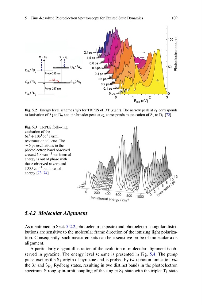
Fig. 5.2 Energy level scheme (left) for TRPES of DT (right). The narrow peak at ε 1 corresponds
to ionisation of S 2 to D 0 and the broader peak at ε 2 corresponds to ionisation of S 1 to D 1 [72]
Fig. 5.3 TRPES following
excitation of the
6a 1 + 10b 1 6b 1 Fermi
resonance in toluene. The
∼ 6 ps oscillations in the
photoelectron band observed
around 500 cm −1 ion internal
energy is out of phase with
those observed at zero and
1000 cm −1 ion internal
energy [73, 74]
5.4.2 Molecular Alignment
As mentioned in Sect. 5.2.2, photoelectron spectra and photoelectron angular distributions are sensitive to the molecular frame direction of the ionizing light polarization. Consequently, such measurements can be a sensitive probe of molecular axis
alignment.
A particularly elegant illustration of the evolution of molecular alignment is observed in pyrazine. The energy level scheme is presented in Fig. 5.4. The pump
pulse excites the S 1 origin of pyrazine and is probed by two-photon ionisation via
the 3s and 3p z Rydberg states, resulting in two distinct bands in the photoelectron
spectrum. Strong spin-orbit coupling of the singlet S 1 state with the triplet T 1 state
109
Fig. 5.2 Energy level scheme (left) for TRPES of DT (right). The narrow peak at ε 1 corresponds
to ionisation of S 2 to D 0 and the broader peak at ε 2 corresponds to ionisation of S 1 to D 1 [72]
Fig. 5.3 TRPES following
excitation of the
6a 1 + 10b 1 6b 1 Fermi
resonance in toluene. The
∼ 6 ps oscillations in the
photoelectron band observed
around 500 cm −1 ion internal
energy is out of phase with
those observed at zero and
1000 cm −1 ion internal
energy [73, 74]
5.4.2 Molecular Alignment
As mentioned in Sect. 5.2.2, photoelectron spectra and photoelectron angular distributions are sensitive to the molecular frame direction of the ionizing light polarization. Consequently, such measurements can be a sensitive probe of molecular axis
alignment.
A particularly elegant illustration of the evolution of molecular alignment is observed in pyrazine. The energy level scheme is presented in Fig. 5.4. The pump
pulse excites the S 1 origin of pyrazine and is probed by two-photon ionisation via
the 3s and 3p z Rydberg states, resulting in two distinct bands in the photoelectron
spectrum. Strong spin-orbit coupling of the singlet S 1 state with the triplet T 1 state